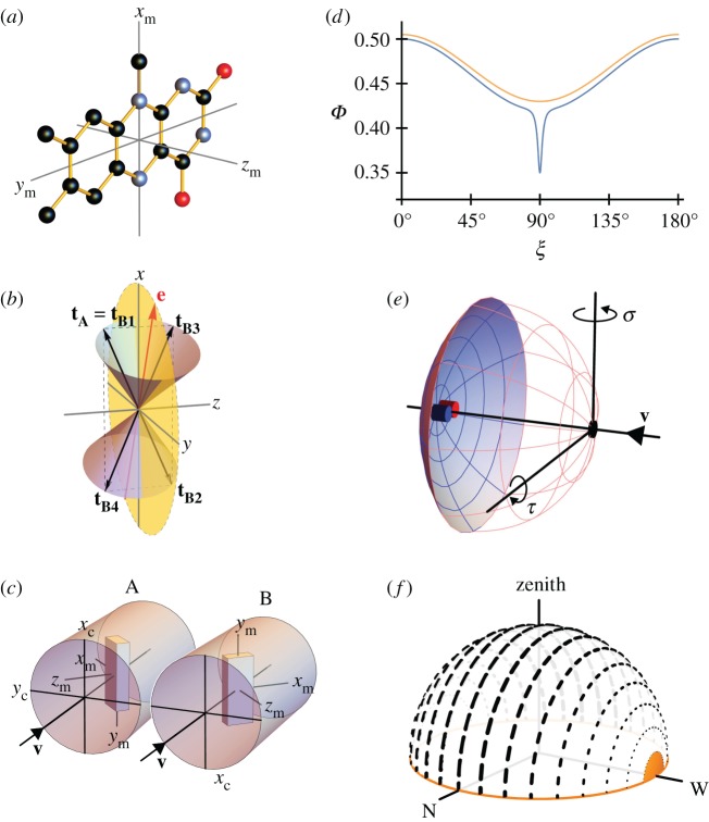
Figure 1.
(a) Structure of the tricyclic flavin component of the flavin adenine dinucleotide chromophore in cryptochrome including the molecular axis system, (xm, ym, zm), used in the text. The transition dipole moment is approximately parallel to ym, the long, in-plane axis of the aromatic ring system. The approximate symmetry axis of the magnetic response of a flavin-containing radical pair is parallel to zm. Black, blue and red balls indicate carbon, nitrogen and oxygen atoms. Hydrogen atoms are not shown. (b) Representation of the transition dipole moment vectors in equations (2.1), (2.2) and (2.4). The incoming light propagates along the z-axis. e is an arbitrarily chosen electric vector (red) in the xy-plane (yellow) and is the axis of the two cones. tA is the TDM of an arbitrarily oriented magnetoreceptor molecule in cell A. For the particular e-vector shown in the figure, any molecule in cell B whose TDM vector points from the origin to any point on the surface of one of the two cones has the same probability of absorbing a photon as does the molecule whose TDM vector is tA (because all such vectors have the same value of cos2ω (equation (2.3)). However, only four of these vectors, tB1, tB2, tB3 and tB4 (all in the xz plane) give the same probability of photon absorption as tA for any direction of e in the xy plane. tB1 is identical to tA. tB2 = −tB1 (i.e. tB1 exactly inverted). tB3 and tB4 are, respectively, the reflections of tB1 and tB2 in the xy plane. Only tB4 is related to tA by a rotation around the z-axis. (c) Schematic representation of two adjacent cylindrical photoreceptor cells, A and B, in the retina. The direction of propagation of the incoming light, v, is parallel to zc, the long symmetry axis of the cells. A and B are identical apart from a 180° rotation around v. Both cells are considered to contain populations of aligned flavin chromophores, one of which is shown in each cell as a cuboid with approximately the same aspect ratios as the flavin structure in (a). The TDM vectors (ym) are parallel in A and B and both are perpendicular to zc. The magnetic symmetry axes (zm) are at 65° to zc and lie in the yczc-plane. (d) Idealized orientation-dependence of the yield of the signalling state of cryptochrome formed from the flavin–tryptophan radical pair state of the protein. Φ(ξ) is modelled (equation (2.5)) so as to resemble the results of calculations presented in Ref. [47] using a0 = 0.5, a1 = −0.08, a128 = −0.01, a512 = −0.03, a2048 = −0.03. ξ is the angle between the magnetic field axis and the zm axis of the flavin structure shown in (a). The orange and blue traces are appropriate for radical pairs with short and long lifetimes, respectively. The former, which includes just the a0 and a1 terms, has been shifted vertically by +0.015 for clarity. (e) Schematic representation of an eye. Cells A and B (small blue and red cylinders) are positioned at the centre of the retina (pink/blue spherical section), diametrically opposite the pupil (black spot). The magnetic responses of A and B are calculated for different ‘lines of sight’ of the eye, obtained by rotating the eye around the indicated axes. σ and τ are the yaw and pitch angles, respectively. (f) Representation of skylight polarization according to the Rayleigh sky model when the sun is on the horizon (orange) in the west. The directions and weights of the short black lines represent the e-vectors of light perceived by an observer at the centre of the celestial hemisphere.

