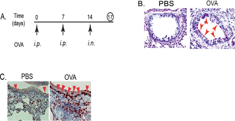
Figure 1. Increased infiltration of MDSCs was accompanied by up-regulated gene expression of KLF4 and TSLP in the lung upon asthmatic challenge.

A. An accute mouse model of allergic asthma as described in the Materials and Methods. i.p. represents intraperitoneal injection and i.n. represents intranasal injection. B. Mouse lung tissues were obtained, cut and mounted on slides, and PAS (Periodic Acid Schiff) staining performed. Red arrows indicated representative stain-positive goblet cells. C. IHC staining of KLF4 from control and OVA-challenged lung tissues using KLF4 antibodies. Red arrows indicate representative KLF4–positive epithelial cells.
