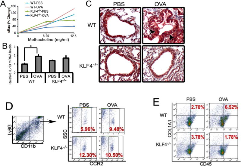
Figure 4. Attenuated AHR and fibrosis in an acute model of allergic asthma in KLF4 knockout mice, accompanied by significantly decreased numbers of fibrocytes.

A. Measurement of specific airway resistance (sRaw) by non-invasive whole-body plethysmography in the WT and KLF4−/− mice upon OVA challenge as described in the Materials and Methods. B. IL-13 expression levels in the lungs of the wild type (WT) and KLF4−/− mice upon control or OVA challenge were assessed by qRT-PCR. n=3, *p<0.05. C. Trichrome staining to examine lung fibrosis before and after OVA challenge in WT and KLF4−/− mice. Arrows point to possible fibrotic areas. D. After gating CD11b+Ly6G+ MDSCs, populations of CCR2+MDSCs in the lungs of WT and KLF4−/− mice before and after OVA challenge were measured by flow cytometry. E. Simiar to D, CD45+COL1A1+ fibrocytes in the lungs were measured by flow cytometry.
