
Figure 2.
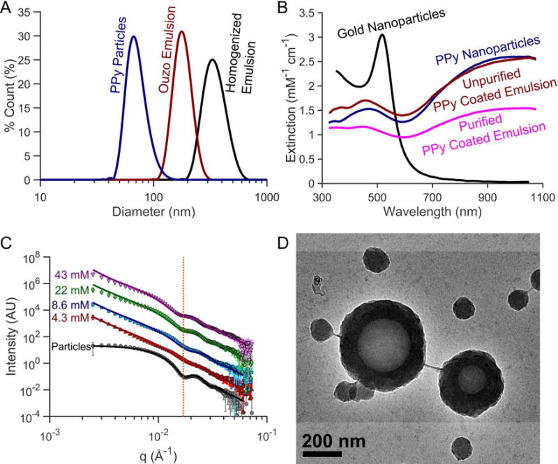
(A) Dynamic light scattering (DLS) particle size distribution, (B) optical extinction spectra, (C) small-angle X-ray scattering (SAXS), and (D) transmission electron microscopy (TEM) image of PPy-coated contrast agents. (A) PPy coated emulsion contrast agents synthesized using high-pressure homogenization produce nanoemulsions between 300–400 nm in diameter. Spontaneously nucleating the droplets for PPy coated emulsion contrast agents can easily produce smaller, monodisperse droplets. The homogenized droplets shown are 357 nm (Polydispersity index (PDI) = 0.102) while the ouzo synthesized droplets were 184 nm (PDI = 0.081) in diameter. (B) PPy-based contrast agents exhibit optical extinction per particle comparable to gold nanoparticles with the added benefit of a broader optical extinction spectrum in the near IR covering the optical diagnostic window for in vivo photoacoustic imaging. (C) SAXS profiles show similar scattering patterns between PPy coated emulsions synthesized with varying pyrrole concentrations, which are also distinctly different from the scattering from solid PPy particles. The line shown in panel C highlights the inflection corresponding to the shell thickness of PPy-coated emulsions. From SAXS data fitting, as the pyrrole concentration reduces from 43mM to 4.3mM, the shell thickness decreases from 41.1 nm to 33.3 nm. SAXS profiles of the particle sample were shifted down two decades while the 8.6, 22, and 43 mM profiles were shifted 1, 2, and 3 decades above the 4.3 mM curve for ease of visualization. The emulsions were isolated from excess particles by centrifuging, removing the supernatant containing the excess particles and resuspending the sedimented emulsion in filtered DI water.