
Figure 5.
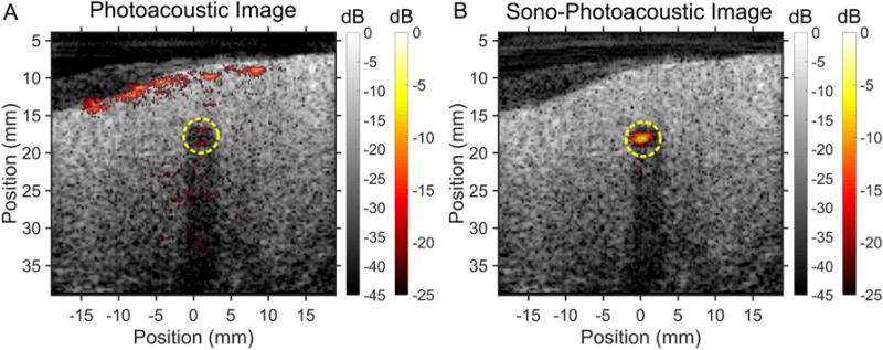
Sequence of images taken using (A) PA imaging of a PA agent versus (B) SPA imaging of PPy coated PFP as the contrast agent in chicken breast tissue at room temperature (25°C). Images were acquired using a surface fluence of 45 mJ/cm2 at λ = 1064 nm and an MI of 0.67 (1.16 MPa). The contrast agents were contained within a 4 mm diameter thin walled plastic tube (yellow dashed line) inserted into the tissue. The tube created an acoustic shadow, seen in the ultrasound images, due to acoustic reflections from the tube wall. Localizing the PA agent is made more difficult due to the PA contrast generated by the tissue itself. In addition to having greater contrast enhancement from the SPA agent during SPA imaging, the PA contrast produced from the tissue is suppressed due to linear PA signal subtraction in the SPA sequence.