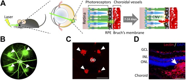
Figure 3.
Laser-induced CNV model. A) Scheme of laser-induced CNV in mice. Adult mice are anesthetized and pupils are dilated. Laser burns are produced in the 3, 6, 9, and 12 o’clock positions around the optic disc (OD), with the laser focused on the RPE. The presence of a subretinal bubble confirms that the laser impact caused the disruption of Bruch’s membrane and RPE, which is necessary for the induction of successful CNV lesions. At 1 and 2 wk after laser treatment, CNVs growing into the subretinal space are observed and analyzed by using fundus fluorescein angiography (FFA) and dissected choroidal flat mounts with isolectin staining. B) A representative image of FFA from a mouse on d 6 after laser burns shows the formation of CNV (green lesions, arrowheads) beneath the retinal vasculature (green). C) A fluorescence microscopy image of an isolectin-stained flat mount of the mouse choroid/RPE at 1 wk after laser treatment shows the extent of CNV lesions (arrowheads). Scale bars, 500 µm. D) Immunohistologic examination of a cross-sectioned mouse eye at 1 wk after laser photocoagulation shows extension of the CNV lesion (red, stained with isolectin; arrow) into the subretinal space between photoreceptors and RPE. Scale bars, 100 µm. GCL, ganglion cell layer; INL, inner nuclear layer; ONL, outer nuclear layer.

