
Figure 4.
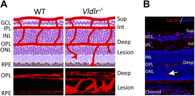
Vldlr−/− mouse model of retinal angiomatous proliferation. A) Schematic diagrams (top) show the retinal neuronal layers (purple) and 3 layers of retinal blood vasculature (superficial, intermediate, and deep; red) on cross-section of wild-type (WT) and Vldlr−/− mice, with blood vessels in Vldlr−/− retinas extending from the deep layer toward RPE. Three-dimensional reconstructed images of vascular lesions in the photoreceptor layer of Vldlr−/− retinas compared with the avascular photoreceptor layer in WT retinas (bottom). Blood vessels were stained with isolectin (red, vessel marker). B) Immunohistochemical staining with isolectin (red) and DAPI (blue, nuclear marker) on a Vldlr−/− mouse retinal cross-section shows abnormal vessel growth (arrows) that originates from the deep vessel layers through the normally avascular photoreceptor layer [outer nuclear layer (ONL)] and toward RPE [reprinted from Hua et al. (112), copyright owned by the Association for Research in Vision and Ophthalmology]. Deep, deep retinal vessel; GCL, ganglion cell layer; INL, inner nuclear layer; Int, intermediate retinal vessels; IPL, inner plexiform layer; OPL, outer plexiform layer; Sup, superficial retinal vessel.