Figure 1.
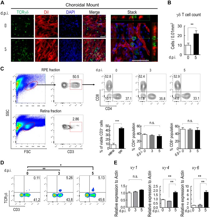
Choroidal γδ–T-cell infiltration after RPE injury. A) Immunostaining of TCRγδ (green) on whole mounts of choroid/sclera prepared from mice that were perfused with endothelial cell dye, Dil (red). Nuclei were counterstained with DAPI (blue). A 3-dimensional reconstructed image was generated from a z-stack scan to show extravascular distribution of γδ T cells (stack). B) Quantification of the average number of γδ T cells per unit area on choroidal whole mount (n = 6). C, D) Flow cytometry analyses of enriched mononuclear cells that were isolated from RPE/choroid or retina for expression of CD3, CD4, CD8 (C) and TCRγδ (D). E) RT-PCR measurement of choroidal γδ–T-cell subtype. γδ T cells were purified by Ab-conjugated magnetic beads at indicated times after NaIO3 treatment. N.s., not significant. Data presented are averages from 3 separate experiments (means ± sem). **P < 0.01. Scale bars, 20 μm.

