Figure 6.
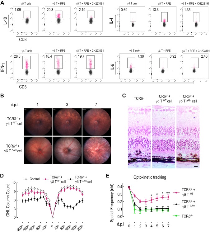
AhR is required for the protective function of γδ T cells against RPE injury. A) Flow cytometry analysis of cytokine expression from γδ T cells that were cocultured with RPE explants in the presence or absence of AhR inhibitor, CH223191. B) Fundus photos from γδ–T-cell–deficient mice that were treated with NaIO3 at indicated times after adoptive transfer of purified γδ T cells from either wild-type or AhR-deficient mice. C) Histopathology of γδ–T-cell–deficient mice after receiving adoptive transfer of wild-type or AhR-deficient γδ T cells and being treated with NaIO3 for 7 d. D, E) Counting of photoreceptor outer nuclei layers in γδ–T-cell–transferred mice (n = 3; D) and visual function examination by optokinetic tracking (n = 5; E). Data are presented as means ± sem. Statistical differences are determined by either 1-way ANOVA with Bonferroni test (D) or Student’s t test (E). *P < 0.05, **P < 0.01. Scale bars, 20 μm.

