Figure 7.
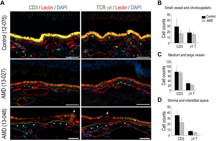
Choroidal γδ T cells in human donor eyes. A) Representative images of immunolabeling of CD3 and γδ TCR on adjacent sections that were prepared from human donor eyes from either patients with AMD or age-matched controls. Vascular endothelium was labeled with lectin (red fluorescence). Arrow indicates drusen; arrowhead indicates areas with choriocapillaris dropout. B–D) Quantification data showing the number of CD3+ cells and γδ T cells in small (B) and larger (C) choroidal vessels and stoma (D) on either control or AMD sections. Scale bars, 50 μm.

