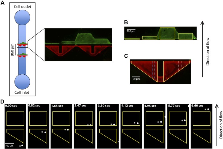
Figure 1.
The microfluidic device showing the distinct E-selectin- and HA-patterned regions on the substrate. A) The E-selectin (red)- and HA (green)-patterned regions separated by a gap distance of 30 µm in the direction of flow in the microfluidic device. B) Four HA patterned regions with lengths (in the direction of flow) of 10, 40, 160, and 80 μm are outlined in yellow. C) 2 E-selectin regions are outlined in yellow. Each region is divided laterally into ten 20-µm-wide sections of decreasing lengths in the direction of flow. Two sections of different lengths are depicted by the white and blue dotted lines. E-selectin and HA patterned regions were identified after labeling with Alexa-488 (green) and Alexa-568 (red), respectively. D) Pa03c pancreatic cancer cell (white asterisks) rolling on E-selectin region and adhering to the HA region (yellow).

