Fig. 4.
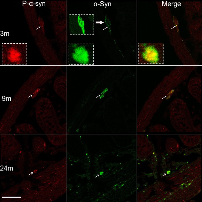
Age-dependent phosphorylation of α-syn in Meissner’s plexus of the small intestine. Profiles of phospho-α-syn (P-α-syn, left), pan-α-syn (middle), and their co-localization (right), in 3, 9, and 24 month-old mice. The photomicrographs acquired under a confocal microscope show increased phosphorylation of α-syn with age (left). Subsets of pan-α-syn-positive profiles (middle) are overlapped with phospho-α-syn-positive profiles (right) (arrows and the insets). Scale bar 50 μm.

