Fig. 6.
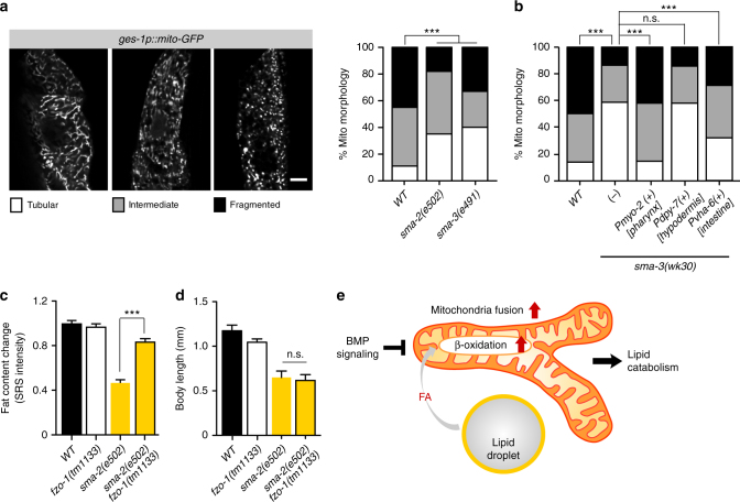
BMP signaling regulates mitochondrial dynamics cell non-autonomously. a Mitochondrial morphology was analyzed using mito-GFP in intestinal cells (zcIs17[ges-1p::mitoGFP]), and classified as tubular, intermediate, and fragmented (representative images are shown, scale bar = 10 µm). The BMP/sma mutants display significantly increased levels of tubular mitochondrial morphology, compared to wild type worms, ***P < 0.001, χ 2 test, n = 100. b Increased levels of tubular mitochondrial morphology in the sma-3 mutant is rescued fully by expressing sma-3 using the myo-2 (pharyngeal muscle) promoter, partially by the vha-6 (intestine) promoter, but not by using the dpy-7 (hypodermis) promoter. ***P < 0.001, n.s. P > 0.05, χ 2 test, n = 100. c The loss-of-function mutant of fzo-1(tm1133) significantly increases the lipid content level of the sma-2 mutant. Error bars represent s.e., ***P < 0.001, Student’s t-test (unpaired, two-tailed), n = 41 for WT, n = 68 for fzo-1, n = 35 for sma-2, n = 74 for fzo-1;sma-2. d The fzo-1 mutant does not rescue the small body size of the sma-2 mutant. Error bars represent s.e., n.s. P > 0.05, Student’s t-test (unpaired, two-tailed), n = 22 for WT, n = 28 for fzo-1, n = 14 for sma-2, and n = 16 for sma-2;fzo-1. e The model of lipid catabolism regulation by BMP signaling. BMP signaling inhibits mitochondrial β-oxidation and mitochondria fusion. When BMP signaling is blocked, the induction of mitochondrial β-oxidation and fusion leads to increased lipid catabolism

