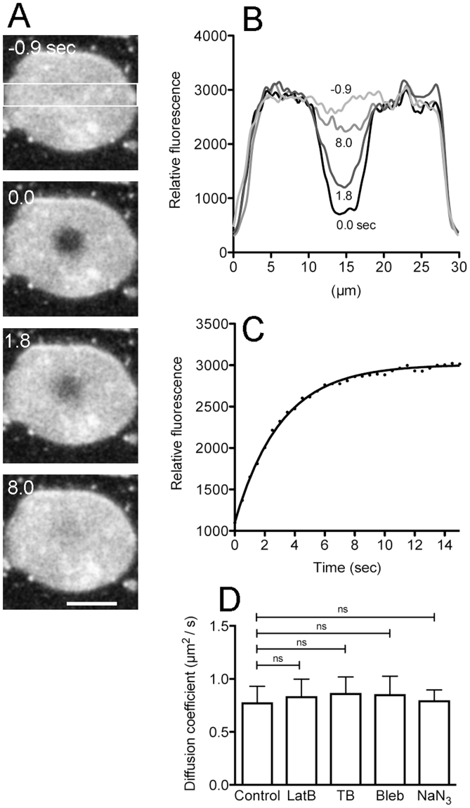
Figure 5.
Lateral diffusion of lipids is not affected by cytoskeletons. The cell membrane was stained with Polaric, a similar fluorescent lipid analogue to CellMask Orange. (A) Typical fluorescence images when a small region of the cell membrane was photobleached (0 sec). (B) The fluorescence intensity profiles in the white rectangle in panel A at each time (−0.9, 0.0, 1.8, and 8.0 sec). Note that the fluorescence recovered by a simple lateral diffusion of the lipids. (C) A time course of fluorescence recovery in the bleached circles. (D) A comparison of the diffusion coefficient in the presence of inhibitors (latrunculin B, thiabendazole, blebbistatin, and sodium azide), indicating that all these inhibitors do not affect the lateral diffusion of the membrane lipids (n = 20 each). Data are presented as mean ± SD and analyzed by one way ANOVA with Tukey’s multiple comparison test. ns, not significant. Bar, 10 µm.

