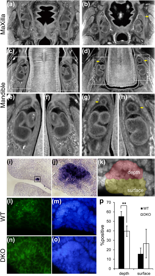
Figure 2.
Formation of the supernumerary tooth in the double KO homozygote of MRCS1 and MFCS4. Transverse sectional micro-CT images of the maxilla (a,b) and mandible (c–h) in the wild type (WT) (a,c,e,f) and the DKO mouse (b,d,g,h). Maxillary tooth in the WT (a) and the DKO mouse (b). Mandibular tooth in the WT (c,e,f) and the DKO mouse (d,g,h). Magnified pictures of the insets of c (e,f) and of d (g,h). Yellow arrows indicate supernumerary teeth. The double KO homozygote of MRCS1 and MFCS4 is schematically illustrated in Supplementary Fig. 1e. Expression of Shh mRNA in the dental placode of the WT at E12.5 (i) and high magnification of the dental placode (j, an open box in i). Nuclei of the dental placode were stained with DAPI. The depth half and the superficial half are shown in red and yellow, respectively (k). Signals for Shh nascent RNA in the dental placode of the WT and DKO (l,n), and cells were stained with DAPI (m,o). The %positive cells transcribing Shh in the depth and the surface of the dental placode (p). The black and open bars depict the wild type and the DKO, respectively. Error bars represent the standard deviations obtained from three independent samples. Two-tailed Student’s t-test was used to test significance of differences in number of signal-positive cells (**P < 0.001).

