Figure 3.
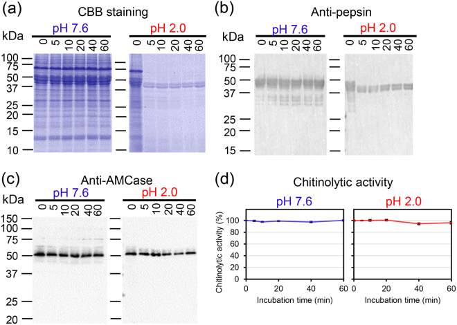
Pepsinogens were converted into active forms and degraded soluble proteins in stomach. Analysis of the endogenous pepsins protease activity in artificially created pig stomach environment (pH 2.0, 37 °C). Soluble protein fractions were prepared from pig stomach tissue in the absence of protease inhibitor and incubated at pH 7.6 or pH 2.0 for up to 60 min and analysed by (a) SDS-PAGE and CBB staining. Full-length gel image is shown in Supplementary Fig. S2. (b) Western blotting using anti-pepsin or (c) AMCase protein using anti-AMCase in the soluble proteins from pig tissue incubated and conducted as described in (a). Full-length blots are shown in Supplementary Fig. S3. (d) AMCase chitinolytic activity in the extract incubated at pH 7.6 or pH 2.0.

