Figure 3.
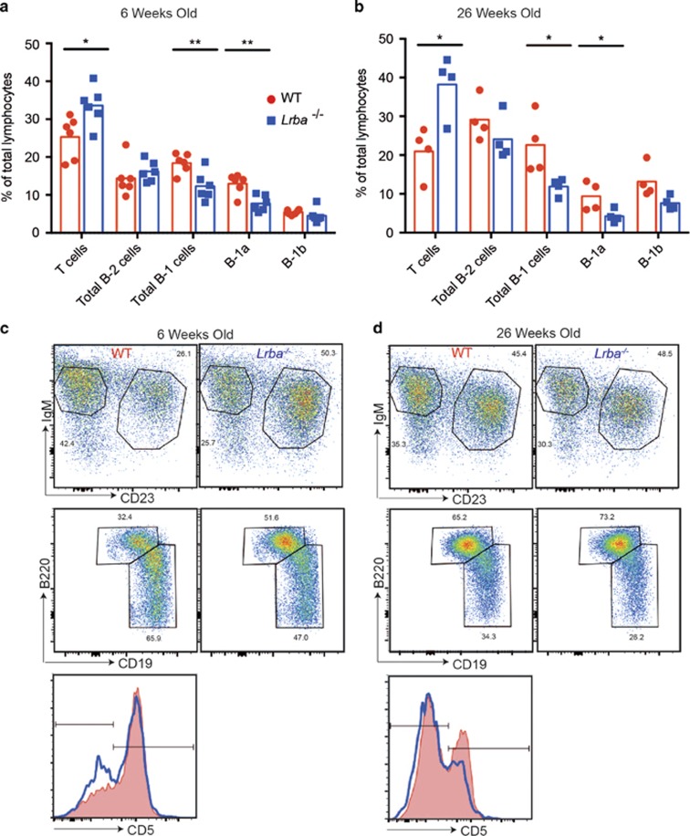
Decreased peritoneal B-1 B cells in LRBA-deficient mice. Flow cytometric analysis of peritoneal cavity lymphocytes from sex-matched WT (red) and Lrba−/− (blue) mice at 6 (n=6) or 26 (n=4) weeks of age, showing individual results and means for each group. (a and b) Percentage of lymphocytes that are T cells (B220− IgM− CD23− CD5+), B-2 cells (CD19−B220hi), B-1 cells (CD19+ B220lo IgM+ CD23−), B-1a cells (CD19+ B220lo IgM+ CD23− CD5+) and B-1b cells (CD19+ B220lo IgM+ CD23− CD5−). (c and d) Representative flow cytometric plots showing gating IgM, CD23, B220 and CD19 staining for B-1 and B-2 cells and representative CD5 histograms of B-1 cells showing gates on CD5+ B-1a and CD5- B-1b cells from mice at 6 (c) and 26 (d) weeks of age. Statistical analysis was carried out using t-test: *P<0.05; **P<0.01. Data are representative of one experiment.

