Figure 3.
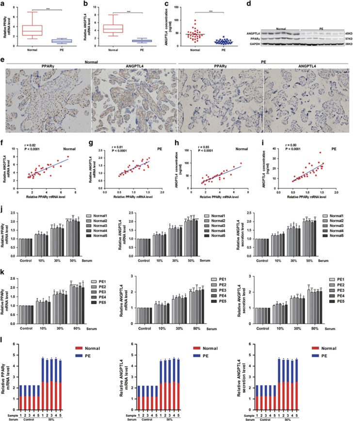
The expression and secretion of ANGPTL4 are decreased and positively correlated with the expression of PPARγ in PE. (a, b) The mRNA levels of PPARγ and ANGPTL4 in placental tissues were detected in PE (n=30) and normal controls (n=30) by qRT-PCR. ***P<0.001 against normal controls. (c) The secretion of ANGPTL4 in the serum was measured among PE (n=30) and normal controls (n=30) by ELISA. ***P<0.001 compared with normal controls. (d) The expression of ANGPTL4 and PPARγ in placental tissues was determined by western blot analysis in seven PE subjects and six normal controls randomly selected from 60 placental tissues. (e) Immunohistochemistry (IHC) was performed to analyse the expression of PPARγ and ANGPTL4 in PE subjects (n=30) and normal controls (n=30). (f–i) The relationship between the mRNA expression of PPARγ and the mRNA and secretion of ANGPTL4 was analysed in PE subjects (n=30) and normal controls (n=30). (j, k) Placental explants were stimulated with serum from PE subjects (n=5) and normal controls (n=5) randomly selected from 60 blood samples. PPARγ and the mRNA and secretion of ANGPTL4 were assessed by qRT-PCR and ELISA. The data are shown as the means±S.E.M. *P<0.05, **P<0.01 and ***P<0.001 compared with corresponding control. (l) The combined data from (j, k) was analysed. The data are shown as the means. *P<0.05 compared with PE

