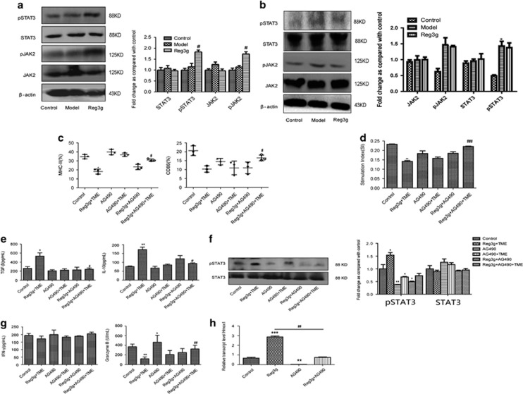
Figure 5.
Reg3g inhibited dendritic cells (DCs) by activating /janus kinase 2/signal transducer and activator of transcription 3 (JAK2/STAT3) signaling pathway. (a) The expressions of phosphorylated (p)STAT3, STAT3, JAK2, and pJAK2 proteins in DCs were analyzed by western blot. (b) The expression of pSTAT3, STAT3, pJAK2, and JAK2 were analyzed by western blot in CD8+ T cells separated using a negative CD8 Isolation Kit (Aubum, CA, USA) from the spleen of control, model, and Reg3g mice. (c and e) DCs were treated with Reg3g+TME, AG490, AG490+TME, Reg3g+AG490, and Reg3g+AG490+TME, respectively. Fluorescence-activated cell sorting (FACS) analysis for the quantitative percentage of CD86 and MHC-II, and the production of interlukin-10 (IL-10) and tumor growth factor-β (TGF-β) in the culture medium were detected by enzyme-linked immunosorbent assay (ELISA). (d) CD8+ T cells were cocultured with DCs at the ratio of 10:1 for 24 h. The proliferation of T cells was then evaluated by the 3-(4,5-dimethylthiazol-2-yl)-2,5-diphenyltetrazolium bromide (MTT) method. (f and g) The protein expressions of STAT3 and pSTAT3 were identified by western blot. The levels of interferon-γ (IFN-γ) and granzyme B in culture medium were estimated by ELISA. (h) The mRNA level of Hmox1 in DCs was analyzed by qRT-PCR. Data were shown as means±S.D. from at least three independent experiments. *P<0.05, **P<0.01, ***P<0.001 compared with the control group; #P<0.05, ##P<0.01, ###P<0.001 compared with the model group (a and b); #P<0.05, ##P<0.01 compared with Reg3g+TME group (c–g)

