Figure 1.
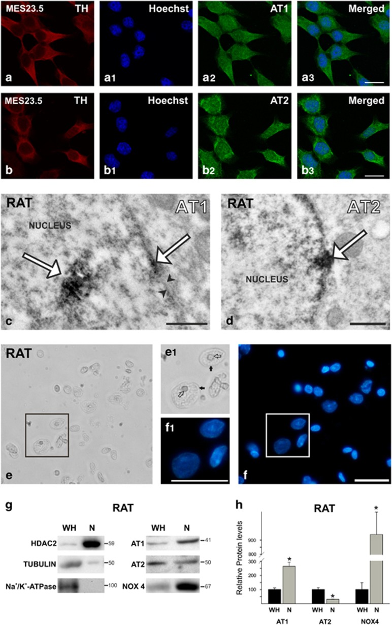
Nuclear AT1, AT2 receptors and NOX4 in nuclei from MES 23.5 dopaminergic neurons and rat nigral region. (a and b) MES 23.5 dopaminergic neurons showing triple immunolabeling for the dopaminergic marker (TH), the nuclear marker (Hoechst), and AT1 (a) or AT2 (b) receptors. (c and d) Electron microscopy of AT1 and AT2 labeling (white arrows) in nuclei and nuclear membranes (between black arrowheads) of rat dopaminergic neurons. (e and f) Nuclei isolated from the rat nigral region in the ventral mesencephalon; the integrity of nuclei was confirmed by microscopic examination with phase contrast (e) and Hoechst staining (f); areas boxed in (e and f) are magnified in (e1 and f1), respectively. (g) WB of whole homogenate (WH) and isolated nuclei (N) from the nigral region showing the expression of AT1 and AT2 receptors, Nox4, as well as different compartment markers used to assess the purity of the nuclei isolation (HDAC2 as a nuclear marker; tubulin as a cytosol marker; and Na+/K+-ATPase as plasma membrane marker). Note the higher expression of AT1 and lower expression of AT2 in the nucleus compared to receptors in WH (g and h). Data are mean±S.E.M. *P<0.05 compared to WH. Student’s t-test (n=3–4). Scale bars: 150 μm (a and b), 0.5 μm (c and d) and 50 μm (e and f)

