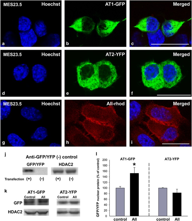
Figure 2.
Presence of fluorescence-tagged angiotensin receptors and fluorescent AII in nuclei. Colocalization (c,f and i) of the fluorescent nuclear marker Hoechst (a,d and g) with AT1-EGFP (b), AT2-YFP (e) or AII-Rhod (h). WB analysis of GFP-/YFP-tagged protein in MES 23.5 transfected (+) and not transfected (−) cells showing the specificity of the common anti-GFP/YFP antibody (j). Treatment of MES 23.5 dopaminergic neuron cell line with AII increased nuclear AT1-EGFP receptor protein but not nuclear AT2-YFP receptor protein relative to nuclei of control cells (k and l). Data are mean±S.E.M. *P<0.05 compared to control. Student’s t-test (n=3–4). AT2-YFP, AT2 tagged to YFP; AT1-EGFP, AT1 tagged to EGFP; AII-Rhod, rhodamine-fluorescent AII. Scale bar: 20 μm (a–i)

