Figure 1.
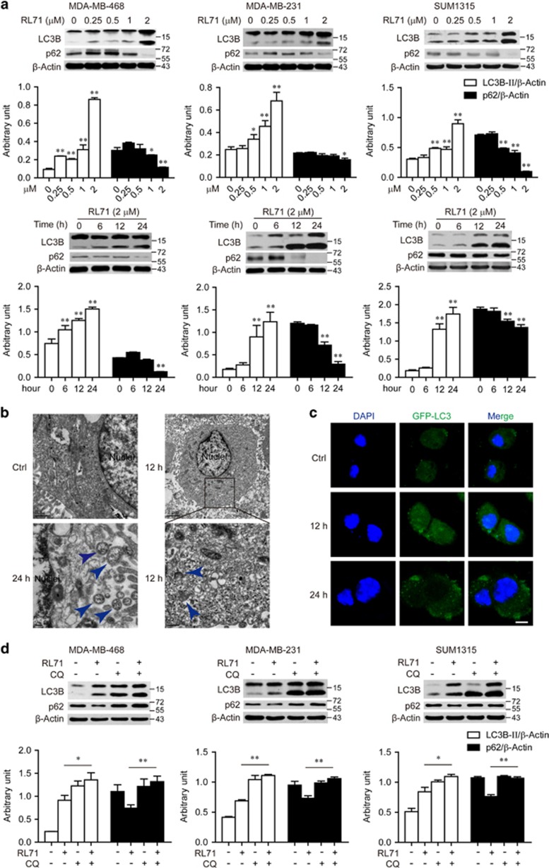
RL71 induces autophagy in TNBC cell lines. (a) Indicated TNBC cell lines were incubated with various concentrations of RL71 (0–2 μM) for 24 h or in the presence of RL71 (2 μM) for different time courses. The protein levels of LC3B and p62 were determined by western blot from whole lysates. β-Actin was used as a loading control. (b) MDA-MB-468 cells were treated with RL71 (2 μM) for 12 or 24 h. Electron microscopy images present the ultrastructure in the cells. Blue arrow, autophagic vaculos with distinct double membrane. (c) MDA-MB-468 cells were transiently transfected with the GFP-LC3 plasmid for 24 h and then treated with RL71 (2 μM) for 12 or 24 h. Representative images show GFP-LC3 localization. Scale bar: 5 μm. (d) Cells were treated with RL71 (1 μM) or CQ (20 μM) alone or in combination for 24 h. The protein levels of LC3B and p62 were determined by western blot. *P<0.05, **P<0.01

