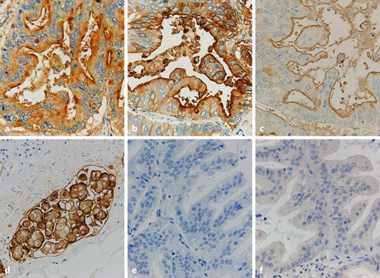
Fig. 3.
Immunohistochemical findings of gastric cancer. The tumor shows diffuse immunopositivity for CA19-9 (a) and CEA (b). The tumor also shows immunopositivity for MUC1 at the luminal surface (c) and occasional immunopositivity in the cytoplasm (d) in the poorly differentiated component in lymphatic vessels. The tumor demonstrates negative immunohistochemical reactions of MUC2 (e) and MUC6 (f). Counterstaining, hematoxylin. Original magnification, ×400.

