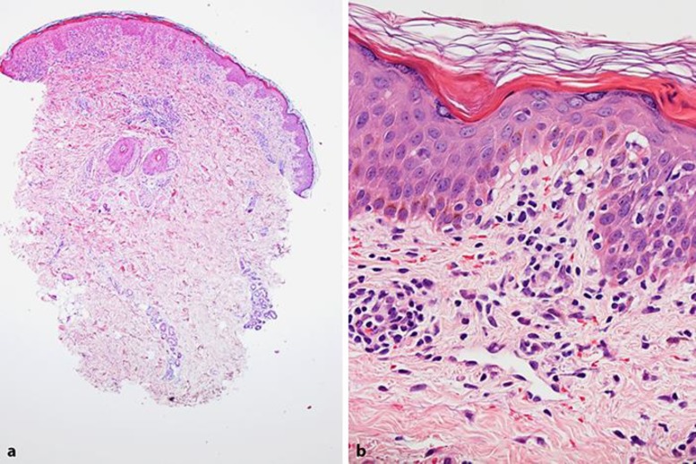
Fig. 2.
Histological features of the reported case. Punch biopsy from the hip showed hyperkeratosis, slight acanthosis, and a superficial lymphocytic infiltrate, most prominent in the papillary dermis. There is perivascular accentuation of the inflammation (HE ×40) (a). The basal keratinocytes show hyperpigmentation and vacuolization, and lymphocytes are found in the epidermis (lichenoid dermatosis). A diffuse and perivascular lymphocytic infiltrate is seen, capillaries are dilated, and there is extravasation of red blood cells (purpura) (HE ×400) (b).

