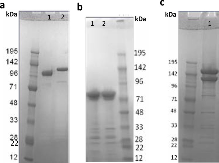
Fig. 1.
SDS-PAGE analysis of SpyTag-DBL1-DBL2x-ID2a as well as CSP-SpyCatcher antigen. a Reduced (lane 1) and non-reduced (lane 2) SDS-PAGE gel of SpyTag-DBL1-DBL2x-ID2a antigen. The band shift indicate the disulfide bond formation, as well as the protein expressed well in baculovirus expression system. On the gel a total of 12 ul sample was loaded, and dilution buffer was used to adjust end concentration into 2 µg. The size of the band correspond to 118 kDa. b Non-reduced (lane 1) and reduced (lane 2) SDS-PAGE gel of CSP-SpyCatcher antigen. No band shift is seen for CSP-SpyCatcher as it contain fewer cycteines for the disulfide bonds as well as band shift formation. The protein expressed as soluble in the baculovirus expression system, and again the dilution buffer was used to adjust end concentration (2 µg) in the total of 12 ul sample loaded on the gel. The theoretical size of CSP-SpyCatcher is 52 kDa but on the SDS-gel it was seen as 72 kDa. C The DBL1-DBL2x-ID2:CSP conjugate formed after overnight incubation (at 4°C) of SpyTag-DBL1-DBL2x-ID2a and CSP-SpyCatcher antigen (lane 1 top). Equal molar amounts of antigens were calculated andmixed (to attain 5 µg final concentration). The excess unconjugated SpyTag-DBL1-DBL2x-ID2a antigen (lane 1 middle).

