Fig. 1.

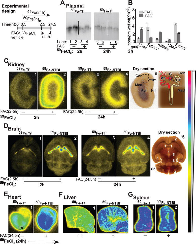
Uptake of 59Fe-NTBI and 59Fe-Tf is organ specific, and 59Fe-NTBI is transported faster than 59Fe-Tf to the brain and the kidney. Experimental design: Time-frame of different treatments for the 2 and 24 h chase experiments in A–G. A) Saturation of plasma Tf with unlabeled FAC results in the distribution of 80% of injected 59Fe to the NTBI pool after 2 h (lanes 1–4). Plasma 59Fe-Tf achieves equilibrium with pools of unlabeled iron in organs by 24 h (lanes 5–8). B) 59Fe-Tf is mainly taken up by the spleen and femur, while 59Fe-NTBI distributes mainly to the liver, kidney, and heart. C) The signal for 59Fe-NTBI in the kidney cortex is 4-fold higher than 59Fe-Tf after 2 h (panels 1 & 2). This difference reduces significantly after 24 h (panels 3 & 4). Both 59Fe-Tf and 59Fe-NTBI show a change in localization from the cortex to the outer medulla after 24 h (panels 3 & 4, panel 5). Panel 5: Dry section used for autoradiography is marked and represented diagrammatically to highlight major sites of iron absorption in the kidney. Cor, cortex; Med, medulla; Pel, pelvis, Hil, hilum; G, glomerulus; PT, proximal tubule; DT, distal tubule. D) The signal from 59Fe-NTBI is significantly higher than 59Fe-Tf in the ventricles and brain parenchyma after 2 h (panels 1 & 2). A significant amount of 59Fe is transported to the brain parenchyma after 24 h, and the signal is higher in 59Fe-NTBI relative to 59Fe-Tf samples (panels 3 & 4, panel 5). Panel 5: Tissue section used for autoradiography is marked to show major sites of iron accumulation in the brain. 1, ventricles; 2, cortex; 3, hippocampus; 4, cerebellar cortex; 5, thalamus; 6, hypothalamus; Cb, cerebellum. E) Accumulation of 59Fe-NTBI in the left ventricular muscle is significantly more than 59Fe-Tf (panels 1 & 2). F) The liver accumulates significantly more 59Fe-NTBI relative to 59Fe-Tf (panels 1 & 2). G) The spleen, on the other hand, accumulates more 59Fe-Tf in comparison to 59Fe-NTBI (panels 1 & 2).
