Fig. 2.

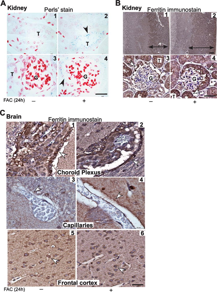
Endothelial and epithelial cells of the brain and kidney upregulate ferritin in iron overloaded mice. A) Perls’ staining of kidney sections shows a blue reaction product of iron in epithelial cells lining the proximal tubules and Bowman’s capsule in FAC treated samples (panels 2 & 4). A weak reaction is also noted in control sections (panels 1 & 3). B) Immunostaining for ferritin followed by enhancement of the reaction with diaminobenzidine shows a much larger area of positivity in the cortex and outer and inner medulla in FAC treated samples relative to controls where the reactivity is limited to the cortex (panels 1 & 2). Glomeruli show a strong reaction for ferritin in cells lining the Bowman’s capsule of FAC treated samples relative to controls (panels 3 & 4). C) Reactivity for ferritin is stronger in brain sections from mice exposed to FAC relative to controls in epithelial cells of the choroid plexus (panels 1 & 2), endothelial cells lining blood capillaries (panels 3 & 4), and neuronal cell bodies in the frontal cortex (panels 5 & 6). Scale bar, 10 μm.
