Figure 2.

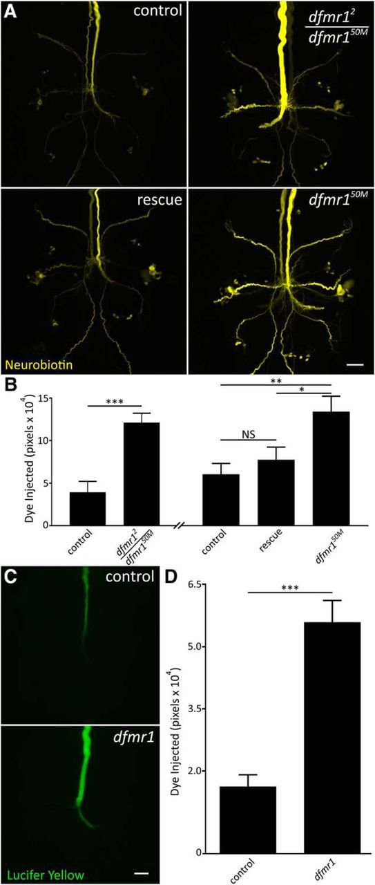
Dye iontophoresis defect is FMRP dependent and charge independent. A, Representative NB images of GFI injections (2 m KAc) for w1118 genetic background (control; top left), heteroallelic dfmr1-null (dfmr12/dfmr150M; top right), wild-type UAS-dFMRP driven with neural elav-Gal4 (rescue; bottom left) in dfmr150M-null background and the dfmr150M-null alone (bottom right). Scale bar, 20 μm. B, Quantification of dye injection in the above four genotypes. The heteroallelic combination and transgenic rescue experiment occurred independently and are displayed with their separate genetic controls. All graphs show data as mean ± SEM. Heteroallelic: control, n = 6; dfmr12/50M, n = 6; rescue control, n = 17; rescue, n = 18; dfmr150M, n = 13. C, Representative images of LY dye injections (in ddH2O) into the GFI neuron in the w1118 genetic background (control; top) and dfmr150M-null mutant (bottom). Scale bar, 20 μm. D, Quantification of LY injection in both genotypes. Control, n = 13; dfmr1 n = 12. Significance determined from two-tailed unpaired t test (B, left; D) and unpaired ANOVA (B, right): *p < 0.05, **p < 0.01, ***p < 0.001.
