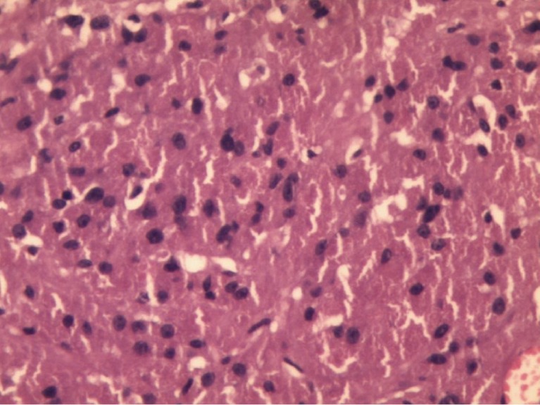
Figure 2.

Microphotograph shows a mesenchymal tumor comprised of diffuse sheets of closely packed large round to polygonal cells containing abundant granular eosinophilic cytoplasm and centrally placed uniform round nuclei (10×).

Microphotograph shows a mesenchymal tumor comprised of diffuse sheets of closely packed large round to polygonal cells containing abundant granular eosinophilic cytoplasm and centrally placed uniform round nuclei (10×).