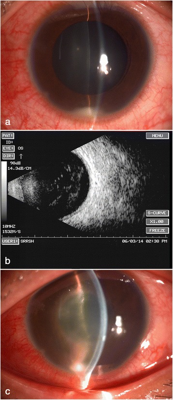
Fig. 1.

Pre-operative evaluation of the infected eye. a slitlamp examination at first presentation revealed moderate congestion in the left eye, with a transparent cornea, anterior cells4+, about two millimeters high hypopyon, a transparent lens. b ultrasonic B scan showed severe vitreous opacity without any posterior vitreous detachment. c after keeping stable for three days with vitreous injection of antibiotics, his left eye deteriorated obviously on the 4th day, with a vision of hand movement and an unobservable fundus
