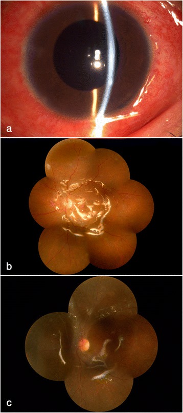
Fig. 2.

Post-operative evaluation of the infected eye. a-b one week after operation, a relatively normal fundus with slight intracameral inflammation was observed, with a best corrected vision of 0.15. c one year later the retina keep attached with slight fibrous proliferation beside optic head and the vision remained 0.1
