
Figure 1.
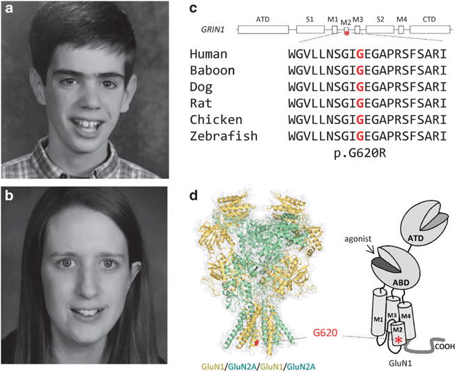
Identification of a GRIN1 missense mutation in patients with developmental delay. (a and b) Facial appearance of Probands 1 and 2. (c) A linear schematic representation of the GluN1 subunit. The position of the mutation is indicated by an asterisk. Gly620 is conserved across vertebral species. ATD, Amino terminal domain; CTD, carboxy terminal domain; M1-4, transmembrane domains (TMs) 1–4; S1 and S2, agonist-binding domains. (d) Homology model of the GluN1/GluN2A receptor built from the GluN2B crystallographic data39 and shown as space fill. The position of the alteration p.G620R is indicated by the red color in region of TM M2.