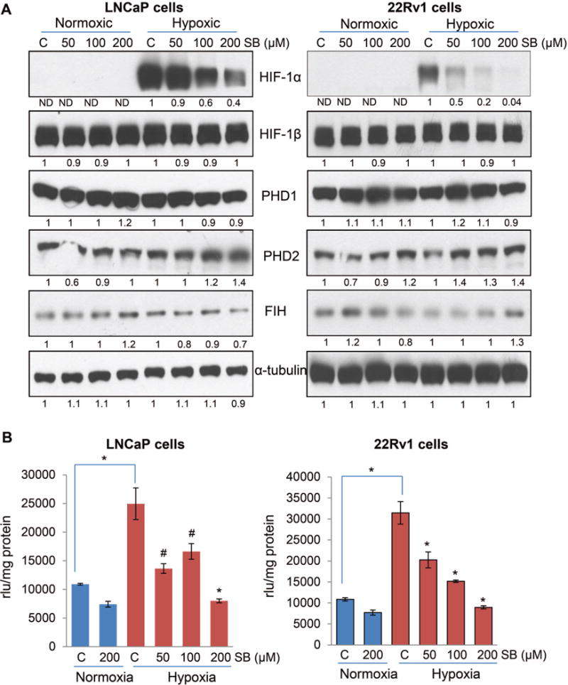
Figure 4.

Silibinin inhibits HIF-1α expression and NOX activity in PCa cells under hypoxic conditions. (A) LNCaP and 22Rv1 cells were cultured under normoxic (21% O2) or hypoxic (1% O2) conditions in the presence of DMSO or silibinin (50–200 μM) for 6 hrs. At the end, total cell lysates were prepared and analyzed for HIF-1α, HIF-1β, FIH, PHD1 and 2 by Western blotting. Membranes were stripped and reprobed for α-tubulin to assess equal protein loading. Densitometry data presented below the bands are ‘fold change’ as compared with respective control after normalization with respective loading control (α-tubulin). ND: Not detectable. (B) LNCaP and 22Rv1 cells (4 × 10⁵ cells/60 mm culture dish) were grown under standard culture condition and after 36 hrs of seeding cells were cultured under normoxic (21% O2) or hypoxic (1% O2) conditions in the presence of DMSO or silibinin for 24 hrs. At the end, cells were harvested and NOX activity was measured as mentioned in ‘Materials and Methods’ and represented as rlu/mg protein. Each value represents mean ± SEM of three samples for each treatment. *, p ≤ 0.001; #, p ≤ 0.01
