
Figure 4.
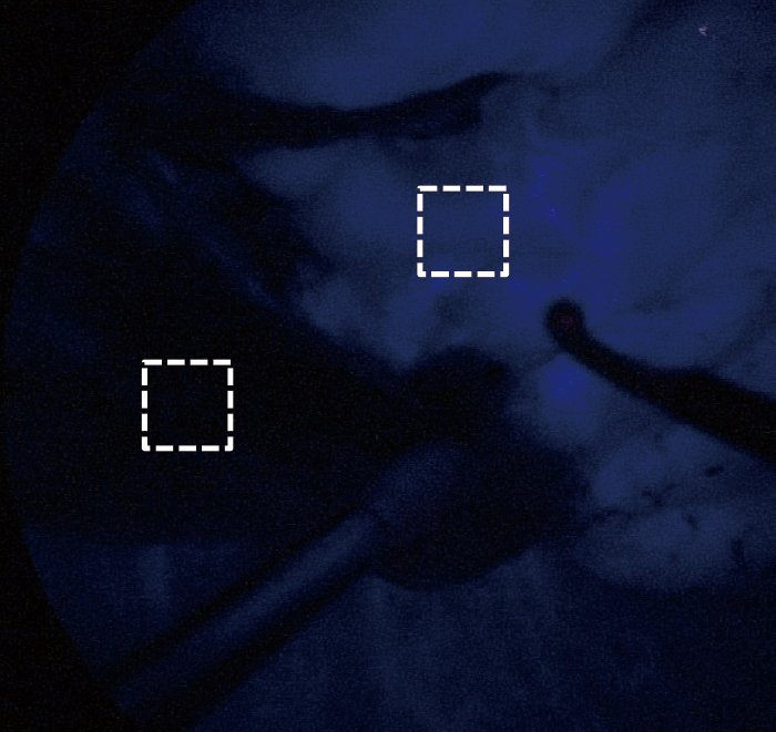
Indocyanine green fluorescence images. Target segment looked dark, and residual segment looked bright. Acquisition of luminosity values using image-analysis software. A 1×1-cm region of interest was identified for target and non-target segments.