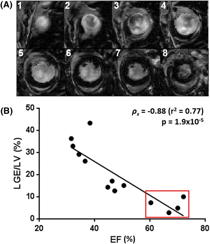
Figure 5.

(A) short‐axis view of late‐gadolinium‐enhanced magnetic resonance imaging (LGE MRI) in an infarcted mouse heart (1, most apical slice; 8, most basal slice). Hyperintensity within the myocardium corresponds to infarcted tissue. (B) correlation of ejection fraction (EF) and area of gadolinium enhancement in the infarcted cohort for the determination of infarct severity. The red box denotes animals with minor cardiac impairment [EF > 50%, total infarct volume as a percentage of the left ventricular volume (LGE/LV) < 10%] where surgery was ineffective. These animals were excluded from subsequent analysis. LV, left ventricle
