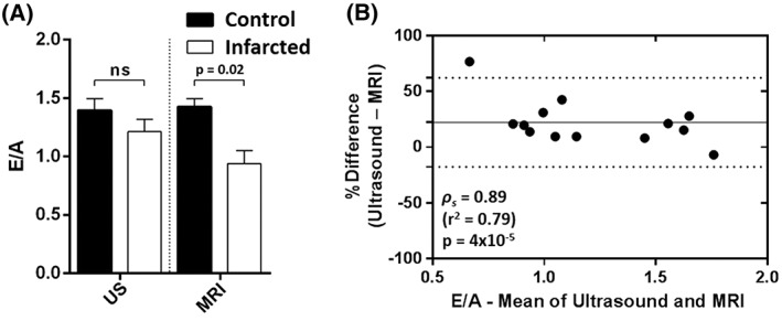
Figure 6.

(A) comparison of early‐filling to atrial/late‐filling ratio (E/A) measurements determined by ultrasound (US) and high‐temporal‐resolution cinematic magnetic resonance imaging (HTR‐CINE MRI) in the infarcted cohort of mice with ejection fraction (EF) < 50% and total infarct volume as a percentage of the left ventricular volume (LGE/LV) > 10% (n = 9). There was a significant difference in E/A between control and infarcted mice when measured with MRI (p = 0.02, Mann–Whitney U‐test), which was not observed with ultrasound (p = 0.15). Ns, not significant. (B) Bland–Altman plot of E/A between ultrasound and MRI across all 13 animals in the infarcted cohort. E/A values tended to be lower when assessed with MRI relative to ultrasound (bias, 22%). Spearman's coefficient showed that the two modalities were strongly correlated (ρ s = 0.89, p = 0.00004)
