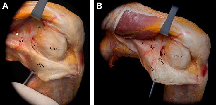
Figure 4.
(A) With further posterior reflection of the superficial iliotibial band (sITB) and blunt separation from the deeper layers, the capsulo-osseous layer (black arrowhead) can be appreciated. The white arrowhead indicates the branches of the superior genicular artery. (B) Proximal, the longitudinally aligned fibers of the intermuscular septum (IS) can be differentiated from the Kaplan fibers (KF). Further, retraction of the sITB reveals the deep ITB (dITB), which merges with the sITB distally. No distinct anterolateral ligament could be observed. The asterisk highlights the accessory insertion of the KF.

