Abstract
At the Abony-Turjányos dűlő site, located in Central Hungary, a rescue excavation was carried out. More than 400 features were excavated and dated to the Protoboleráz horizon, at the beginning of the Late Copper Age in the Carpathian Basin, between 3780–3650 cal BC. Besides the domestic and economic units, there were two special areas, with nine-nine pits that differed from the other archaeological features of the site. In the northern pit group seven pits contained human remains belonging to 48 individuals. Some of them were buried carefully, while others were thrown into the pits. The aim of this study is to present the results of the paleopathological and molecular analysis of human remains from this Late Copper Age site. The ratio of neonates to adults was high, 33.3%. Examination of the skeletons revealed a large number of pathological cases, enabling reconstruction of the health profile of the buried individuals. Based on the appearance and frequency of healed ante- and peri mortem trauma, inter-personal (intra-group) violence was characteristic in the Abony Late Copper Age population. However other traces of paleopathology were observed on the bones that appear not to have been caused by warfare or inter-group violence. The remains of one individual demonstrated a rare set of bone lesions that indicate the possible presence of leprosy (Hansen’s disease). The most characteristic lesions occurred on the bones of the face, including erosion of the nasal aperture, atrophy of the anterior nasal spine, inflammation of the nasal bone and porosity on both the maxilla and the bones of the lower legs. In a further four cases, leprosy infection is suspected but other infections cannot be excluded. The morphologically diagnosed possible leprosy case significantly modifies our knowledge about the timescale and geographic spread of this specific infectious disease. However, it is not possible to determine the potential connections between the cases of possible leprosy and the special burial circumstances.
Introduction
Archaeological background
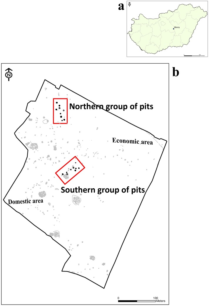
The Abony-Turjányos dűlő site (Fig 1a) is located in central Hungary, where a rescue excavation was carried out between 2004 and 2008. In the site more than 400 features were excavated and dated to the ProtoBoleráz horizon, immediately before the Baden cultural complex of the Late Copper Age in the Carpathian Basin [1–5]. The Accelerator Mass Spectroscopy (AMS) radiocarbon dating indicates that the settlement was used between 3780 and 3650 cal BC. The measurements was made at the Vienna Environmental Research Accelerator (VERA) on samples of animal and human bones [5].
Fig 1.
a: Location of Abony-Turjányos dűlő site. b: Site map of Abony-Turjányos dűlő.
In the southern and southwestern part of the site different kinds of pits—such as storage pits, refuse pits and pit complexes—were unearthed that contained large numbers of collapsed vessels (Fig 1b). The pits also yielded raw materials for housing and a great amount of daub rubble. Some of the daubs were found with imprints of wooden stick and boards, which seem to have belonged to the structure of former houses with wattle and daub walls. These remains indicated that this were the domestic part of the settlement. The eastern and northeastern part of the site was the economic area of the settlement. The excavators assumed that the elongated pits excavated here may have served some function other than storage or clay extraction so could have been used in a special work process [5].
In the economic unit of the settlement there were two special (a northern and southern) area, each with pits that were different in all aspects from the other archaeological features of the site. In the northern group seven pits contained human remains belonging to 48 individuals. Some of them were buried carefully, while others had been thrown into the pits. The skeletons were in clearly separated layers. The pits contained black humus soil and also sterile loess layers. In the rest of these sacrificial pits (bothroi), beside the human burials, animal bones, intact ceramic vessels and sherds were deposited in layers, which suggest the regular repetition of burial activities. The southern group with nine pits was recovered later, but contained no human remains [5].
Other mass graves or multiple burials dating from the Late Copper Age have been found within settlements from the Carpathian Basin, including: Palotabozsok [6,7] Tikos-Homokgödrök [8], Balatonszemes-Szemesi Berek [6,7,9], Balatonőszöd-Temetői dűlő [7,10], Balatonboglár [7,11] and Kaposújlak-Várdomb [7,12]. In the archaeological literature these pits are usually described as sacrificial burials, ritual murders or it is suggested they were connected to warfare or to an epidemic that resulted in the death of several individuals almost simultaneously [6,7,13]. The origin of the Late Copper Age Abony and the other few known contemporary adjacent populations are almost unknown, due to the lack of comparative physical anthropological, ancient DNA and stable isotope data about earlier and contemporary human groups.
During the past two decades, archaeological activity in Hungary has furnished a large number of new cases with paleopathological markers. At the same time, the intensive study of these specific infectious diseases and the development of molecular diagnostic methods has increased the number of publications in this field [14–24].
The aim of this study is to present the results of the paleopathological and molecular analysis of human remains from the Abony Late Copper Age site.
Methods
Paleopathological analysis
The estimation of age-at-death and the sex determination were based on the methods commonly used in physical anthropology [25–32]. The paleopathological lesions were investigated macroscopically [33–35]. Due to the possible ritual function of the pits, there was particular emphasis on any paleopathological alterations that might indicate inter- or intragroup violence, or the presence of infections, such as endocranial lesions, periosteal appositions and porotic hyperostosis [36–40]. Special attention was paid to porotic hyperostosis that can be the sign of a weakened immune system. However, hematological disorders can also indicate an increased susceptibility to infections [41]. Porotic hyperostosis was assessed using a 4-grade-scale: no bone lesion; porotic, cribrotic and trabecular forms [42].
The analysis was supplemented by the investigation of possible signs of ante- and peri- mortem trauma, which can indicate the lifestyle and may also shed light on the circumstances of the death of these individuals. In the trauma analysis, bones were analysed according to Lovell [43], Ortner [35] and Rodríguez-Martín [44].
Computed tomography (CT) examinations were performed using a Siemens Somatom Definition Flash DSCT 2x128 scanner (Siemens AG, Erlangen, Germany). Primary images were obtained using a slice thickness of 0.4 mm. Secondary multiplanar and volume rendering technique (VRT) reconstructions were performed for better visualisation.
Molecular examination for Mycobacterium leprae aDNA
DNA extraction
The recommended protocols of ancient DNA (aDNA) work [45] were followed, with separate rooms for different stages of the process. Bone scrapings were taken from the following four specimens: feature 257 S20 –inferior nasal concha; feature 263 S25 –hard palate; feature 263 S29 –maxillary alveolar process; and feature 263 S36 –inferior nasal spine. Scrapings were crushed in a sterile pestle and mortar. Next, 50–70 mg of each sample was transferred into a 2ml screw-capped Eppendorf tube containing demineralisation solution (EDTA/Proteinase K). Tubes were incubated in a 56°C heating block and mixed daily for 3 days when the samples were solubilised [15]. Samples were then divided and one aliquot treated with 40μl of 0.1 mol−1 of N-phenacylthiozolium bromide (PTB), to cleave any covalent cross-links thus enabling DNA strand separation and amplification [46]. To complete disaggregation of the samples the 2ml tube contents were transferred into 9ml tubes of NucliSens® (bioMérieux) lysis buffer containing 5 mol-1 guanidium thiocyanate solution, and incubated at 56°C. Negative extraction controls were processed in parallel with sample tubes.
Tubes were placed on a rotator for 4 hours, snap-frozen in liquid nitrogen and thawed in a 65°C waterbath. This was repeated twice. Sample were spun at 5000g for 15 mins at 5°C and the supernatants carefully removed into clean, sterile tubes. To capture DNA the supernatants were mixed with 50μl silica suspension (NucliSens®) and centrifuged. The silica pellets were washed once with wash buffer (NucliSens®), twice with 70% (v/v) ethanol (-20°C) and once with acetone (-20°C). After drying in a heating block, DNA was eluted from the silica using 60μl elution buffer (NucliSens®), aliquoted and used immediately or stored at -20°C. Silica supernates (500μl) from PTB-negative samples were also collected from the 9ml tubes of lysis buffer, and the 2.0 ml screw-capped Eppendorf tubes used to wash the silica. After chilling at 5°C, supernates were mixed vigorously for 20 s with 200μl of Protein Precipitation Solution (SLS Ltd., UK) and centrifuged for 3min at 10,000g. Any pellet was discarded and 600μl isopropanol (−20°C) added to 550μl of each supernate. Tubes were mixed by inversion 50 times and spun 3min. Supernates were discarded and tubes washed once with 500μl 70% ethanol (−20°C). After draining, tubes were dried in a heating block. Any precipitated DNA was re-hydrated with 60μl elution buffer (NucliSens®), aliquoted and used immediately or stored at −20°C. Negative extraction controls were processed in parallel with the test samples.
Two repeat batches of DNA extractions were performed subsequently, using 53–65 mg and 45–55 mg of crushed scrapings as described above.
DNA amplification and detection
Initially M. leprae DNA was sought by targeting a specific region of the repetitive element RLEP (37 copies/cell). A two-tube nested PCR was used which give an outer product of 129 base pairs (bp) and a nested PCR product of 99 bp [47]. A hot-start Taq polymerase was used to minimise non-specific primer and template binding. Negative DNA extraction and PCR controls were processed alongside the test sample. Detection of PCR product was by agarose gel electrophoresis, stained with ethidium bromide and visualised under ultraviolet light. As aDNA is normally fragmented, a further pair of RLEP primers with a target region of 111bp was also used in a single-stage PCR reaction [48]. As there are reports of leprosy co-infections with tuberculosis, the first batch of DNA extracts were also examined for the presence of M. tuberculosis aDNA using IS6110 primers P1 and P2 followed by a nested reaction with primers IS3 and IS4, with target sequences of 123 bp and 92 bp respectively [15]. Subsequently a second specific region of the repetitive element RepLep (15 copies/cell) was also examined, and specific primers and probes were used to enable shorter PCR fragments to be detected in a real-time PCR reactions using both the RLEP and RepLep target regions (S1 Table). The PCR probe mix included 2mM bovine serum albumin to reduce PCR inhibition [49,50], 2.0mM MgCl2 and annealing was at 60°C. A hot-start Taq polymerase was used to minimize non-specific primer and template binding. Negative DNA extraction and PCR controls were processed alongside the test sample. Amplification was performed in a final volume of 25μl using a RotorGene 3000 (Qiagen) real-time platform [51].
Material
During the physical anthropological work the analysed human remains were housed in the Institute of Archaeology, Research Centre for the Humanities and later in the Hungarian National Museum. After the archaeological, anthropological and molecular examinations the material will be stored in the Ferenczy Museum Center. The feature and stratigraphic unit numbers of the investigated skeletons are in the S2 Table.
Results
Age and sex distribution
During the physical anthropological work we investigated the remains of 48 individuals (Table 1, S2 Table). In the nine pits differences were observed in the number of buried or thrown-in individuals. In two pits there were no human remains (features 248 and 255), while in feature 263 the skeletal remains of 23 individuals were found. In features 247, 249 and 253 there were only fetuses and neonates. There were inhumations of a female and three neonates in feature 251 (S1 Video) and of one male and one child in feature 250. Most of the individuals were buried in feature 257 (9 skeletons—two females, two males, four subadults and one undeterminable adult—S2 Video) and feature 263 (23 skeletons– 10 females, 7 males, 6 subadults).
Table 1. The sex and age distribution of the buried individuals.
| Age groups | Sex | |||
|---|---|---|---|---|
| Not known | Males | Females | Total | |
| Fetus, Neonate, 0–1 years-old | 16 | – | – | 16 |
| Infans I. (1–7 years-old) | 6 | – | – | 6 |
| Infans II. (8–14 years-old) | 2 | – | – | 2 |
| Juvenile (15–23 years-old) | – | 1 | – | 1 |
| Adult (24–39 years-old) | 1 | 5 | 9 | 15 |
| Mature (40–59 years-old) | – | 4 | 4 | 8 |
| Senile (60+ years-old) | – | – | – | 0 |
| Total | 25 | 10 | 13 | 48 |
Regarding age distribution, the ratio of children, especially with an-age-at death of 0–1 years was high (33.3%). Furthermore, the proportions of juveniles and young adults were low (Table 1).
Ante and peri-mortem trauma
In our previous study we published the preliminary observations of traumatic lesions in the Abony population [52]. In this recent paper we provided a more detailed analysis of trauma on the human bones of 48 individuals (25 subadults and 23 adults). The results are described in S1 Text, S3 and S4 Tables, S1–S9 Figs.
Endocranial lesions
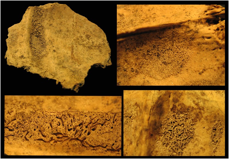
Of all the paleopathology that could be detected, there was only one adult case of endocranial lesions (on a left parietal bone of a female), while these were much more frequent amongst children (Table 2). These usually affected several cranial bones in one individual, mainly the parietals and the occipital bone. From the 18 subadult skulls, endocranial lesions were detected in five cases.
Table 2. The occurence of endocranial lesions.
| Bone | Sub-adults | Adults | Total | ||||||||||||
|---|---|---|---|---|---|---|---|---|---|---|---|---|---|---|---|
| N1 | % | N2 | % | N1+2 | N1 | % | N2 | % | N1+2 | N1 | % | N2 | % | N1+2 | |
| Frontal | 2 | 11.1 | 16 | 88.9 | 18 | 0 | 0.0 | 23 | 100.0 | 23 | 2 | 4.4 | 43 | 96.6 | 45 |
| Left parietal | 0 | 0.0 | 18 | 100.0 | 18 | 1 | 3.6 | 22 | 96.4 | 23 | 1 | 2.2 | 45 | 97.8 | 46 |
| Right parietal | 1 | 5.9 | 16 | 94.1 | 17 | 0 | 0.0 | 23 | 100.0 | 23 | 1 | 2.4 | 41 | 97.6 | 42 |
| Left temporal | 0 | 0.0 | 16 | 100.0 | 16 | 0 | 0.0 | 23 | 100.0 | 23 | 0 | 0.0 | 40 | 100.0 | 40 |
| Right temporal | 0 | 0.0 | 16 | 100.0 | 16 | 0 | 0.0 | 23 | 100.0 | 23 | 0 | 0.0 | 41 | 100.0 | 41 |
| Occipital | 5 | 38.5 | 8 | 61.5 | 13 | 0 | 0.0 | 23 | 100.0 | 23 | 3 | 7.9 | 33 | 92.1 | 38 |
N1 = Yes, N2 = No, N1+2 = Sum of the examinable cases
In two cases the lesions were present on more bones (feature 257 S17: frontal and occipital bones; feature 263 S38 –Fig 2: frontal, right parietal and occipital bones). In three cases the occipital bones were affected (feature 250 S5; feature 257 S18; feature 263 S27).
Fig 2. Endocranial patterns on the frontal and occipital bones, feature 263 S38.
Periosteal lesions
Periosteal lesions can arise from different causes [53,54], but one of the most common is infection [35,55]. Among children periostitis occurs most frequently on the femur and tibia, while among the adults it is mainly on the tibia, the fibula and/or the femur (Table 3). Disaggregated by sex it occured in almost equal proportions, but slightly more frequent among males (Table 4).
Table 3. The occurence of periostitis.
| Sub-adults | Adults | Total | |||||||||||||
|---|---|---|---|---|---|---|---|---|---|---|---|---|---|---|---|
| N1 | % | N2 | % | N1+2 | N1 | % | N2 | % | N1+2 | N1 | % | N2 | % | N1+2 | |
| Left clavicle | 1 | 9.1 | 10 | 90.9 | 11 | 0 | 0.0 | 12 | 100.0 | 12 | 1 | 4.3 | 22 | 95.7 | 23 |
| Right clavicle | 0 | 0.0 | 7 | 100.0 | 7 | 1 | 10.0 | 9 | 90.0 | 10 | 1 | 5.9 | 16 | 94.1 | 17 |
| Left humerus | 1 | 5.3 | 18 | 94.7 | 19 | 1 | 4.8 | 20 | 95.2 | 21 | 2 | 5.0 | 38 | 95.0 | 40 |
| Right humerus | 0 | 0.0 | 16 | 100.0 | 16 | 1 | 5.0 | 19 | 95.0 | 20 | 1 | 2.8 | 35 | 97.2 | 36 |
| Left radius | 0 | 0.0 | 9 | 100.0 | 9 | 1 | 5.3 | 18 | 94.7 | 19 | 1 | 3.6 | 27 | 96.4 | 28 |
| Right radius | 0 | 0.0 | 8 | 100.0 | 8 | 1 | 5.6 | 17 | 94.4 | 18 | 1 | 3.8 | 25 | 96.2 | 26 |
| Left ulna | 1 | 7.7 | 12 | 92.3 | 13 | 1 | 5.6 | 17 | 94.4 | 18 | 2 | 6.5 | 29 | 93.5 | 31 |
| Right ulna | 0 | 0.0 | 12 | 100.0 | 12 | 1 | 5.6 | 17 | 94.4 | 18 | 1 | 3.3 | 29 | 96.7 | 30 |
| Left femur | 2 | 13.3 | 13 | 86.7 | 15 | 5 | 20.8 | 19 | 79.2 | 24 | 7 | 17.9 | 32 | 82.1 | 39 |
| Right femur | 2 | 11.1 | 16 | 88.9 | 18 | 5 | 19.2 | 21 | 80.8 | 26 | 7 | 15.9 | 37 | 84.1 | 44 |
| Left tibia | 2 | 16.7 | 10 | 83.3 | 12 | 10 | 41.7 | 14 | 58.3 | 24 | 12 | 33.3 | 24 | 66.7 | 36 |
| Right tibia | 5 | 33.3 | 10 | 66.7 | 15 | 10 | 40.0 | 15 | 60.0 | 25 | 15 | 37.5 | 25 | 62.5 | 40 |
| Left fibula | 0 | 0.0 | 8 | 100.0 | 8 | 8 | 38.1 | 13 | 61.9 | 21 | 8 | 27.6 | 21 | 72.4 | 29 |
| Right fibula | 0 | 0.0 | 7 | 100.0 | 7 | 6 | 28.6 | 15 | 71.4 | 21 | 6 | 21.4 | 22 | 78.6 | 28 |
N1 = Yes, N2 = No, N1+2 = Sum of the examinable cases
Table 4. The occurence of periostitis according to sex.
| Bones | Males | Females | ||||||||
|---|---|---|---|---|---|---|---|---|---|---|
| N1 | % | N2 | % | N1+N2 | N1 | % | N2 | % | N1+N2 | |
| Left clavicle | 0 | 0.0 | 2 | 100.0 | 2 | 0 | 0.0 | 9 | 100.0 | 9 |
| Right clavicle | 0 | 0.0 | 2 | 100.0 | 2 | 1 | 12.5 | 7 | 87.5 | 8 |
| Left humerus | 1 | 16.7 | 5 | 83.3 | 6 | 0 | 0.0 | 8 | 100.0 | 8 |
| Right humerus | 1 | 16.7 | 5 | 83.3 | 6 | 0 | 0.0 | 9 | 100.0 | 9 |
| Left radius | 1 | 16.7 | 5 | 83.3 | 6 | 0 | 0.0 | 9 | 100.0 | 9 |
| Right radius | 1 | 16.7 | 5 | 83.3 | 6 | 0 | 0.0 | 8 | 100.0 | 8 |
| Left ulna | 1 | 16.7 | 5 | 83.3 | 6 | 0 | 0.0 | 9 | 100.0 | 9 |
| Right ulna | 1 | 16.7 | 5 | 83.3 | 6 | 0 | 0.0 | 7 | 100.0 | 7 |
| Left femur | 2 | 25.0 | 6 | 75.0 | 8 | 2 | 20.0 | 8 | 80.0 | 10 |
| Right femur | 2 | 28.6 | 5 | 71.4 | 7 | 2 | 18.2 | 9 | 81.8 | 11 |
| Left tibia | 3 | 42.8 | 4 | 57.2 | 7 | 5 | 55.6 | 4 | 44.4 | 9 |
| Right tibia | 4 | 57.2 | 3 | 42.8 | 7 | 4 | 44.4 | 5 | 55.6 | 9 |
| Left fibula | 4 | 57.2 | 3 | 42.8 | 7 | 3 | 33.3 | 6 | 66.7 | 9 |
| Right fibula | 3 | 50.0 | 3 | 50.0 | 6 | 3 | 33.3 | 6 | 66.7 | 9 |
N1 = Yes, N2 = No, N1+2 = Sum of the examinable cases
Porotic hyperostosis
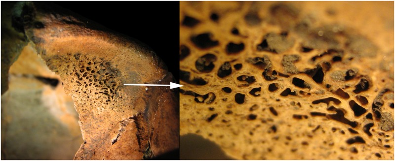
Porotic hyperostosis, suggesting susceptibility for infections, a weakened immune system or haematological disorders [35], was infrequent in this population (Tables 5 and 6). Among the examinable 10 subadults we detected porotic hyperostosis in two individuals. In the 23 adult skulls it was visible in four individuals. Most of them occurred in the orbital roof (cribra orbitalia) (Fig 3) and only once on the occipital bone (cribra cranii) (Table 5). This alteration affected mainly females, which corresponds to the paleopathological literature (Table 6) [55,56].
Table 5. The occurence of porotic hyperostosis.
| Bones | 0 | % | 1 | % | 2 | % | 3 | % | ∑ | |
|---|---|---|---|---|---|---|---|---|---|---|
| Sub-adults | Left orbit | 5 | 62.5 | 2 | 25.0 | 1 | 12.5 | 0 | 0.0 | 8 |
| Right orbit | 7 | 70.0 | 1 | 10.0 | 2 | 20.0 | 0 | 0.0 | 10 | |
| Left parietal bone | 9 | 90.0 | 0 | 0.0 | 1 | 10.0 | 0 | 0.0 | 10 | |
| Right parietal bone | 9 | 100.0 | 0 | 0.0 | 0 | 0.0 | 0 | 0.0 | 9 | |
| Occipital bone | 7 | 100.0 | 0 | 0.0 | 0 | 0.0 | 0 | 0.0 | 7 | |
| Adults | Left orbit | 16 | 72.7 | 4 | 18.2 | 2 | 9.1 | 0 | 0.0 | 22 |
| Right orbit | 19 | 86.4 | 1 | 4.5 | 2 | 9.1 | 0 | 0.0 | 22 | |
| Left parietal bone | 23 | 100.0 | 0 | 0.0 | 0 | 0.0 | 0 | 0.0 | 23 | |
| Right parietal bone | 24 | 100.0 | 0 | 0.0 | 0 | 0.0 | 0 | 0.0 | 24 | |
| Occipital bone | 22 | 95.7 | 1 | 4.3 | 0 | 0.0 | 0 | 0.0 | 23 | |
| Total | Left orbit | 21 | 70.0 | 6 | 20.0 | 3 | 10.0 | 0 | 0.0 | 30 |
| Right orbit | 26 | 81.2 | 2 | 6.3 | 4 | 12.5 | 0 | 0.0 | 32 | |
| Left parietal bone | 32 | 97.0 | 0 | 0.0 | 1 | 3.0 | 0 | 0.0 | 33 | |
| Right parietal bone | 33 | 100.0 | 0 | 0.0 | 0 | 0.0 | 0 | 0.0 | 33 | |
| Occipital bone | 29 | 96.7 | 1 | 3.3 | 0 | 0.0 | 0 | 0.0 | 30 |
0 = not present, 1 = porotic grade, 2 = cribrotic grade, 3 = trabecular grade
Table 6. The occurence of porotic hyperostosis according to sex.
| Grade | Males | Females | Sum | |||
|---|---|---|---|---|---|---|
| N | % | N | % | N | ||
| Left orbit | 0 | 4 | 57.14 | 9 | 75.0 | 13 |
| 1 | 1 | 14.29 | 3 | 25.0 | 4 | |
| 2 | 2 | 28.57 | 0 | 0.0 | 2 | |
| 3 | 0 | 0.00 | 0 | 0.0 | 0 | |
| 100.0 | 100.0 | |||||
| Right orbit | 0 | 4 | 66.7 | 11 | 91.7 | 15 |
| 1 | 0 | 0.0 | 1 | 8.3 | 1 | |
| 2 | 2 | 33.3 | 0 | 0.0 | 2 | |
| 3 | 0 | 0.0 | 0 | 0.0 | 0 | |
| 100.0 | 100.0 | |||||
| Left parietale | 0 | 7 | 100.0 | 12 | 100.0 | 19 |
| 1 | 0 | 0.0 | 0 | 0.0 | 0 | |
| 2 | 0 | 0.0 | 0 | 0.0 | 0 | |
| 3 | 0 | 0.0 | 0 | 0.0 | 0 | |
| 100.0 | 100. | |||||
| Right parietale | 0 | 7 | 100.0 | 13 | 100.0 | 20 |
| 1 | 0 | 0.0 | 0 | 0.0 | 0 | |
| 2 | 0 | 0.0 | 0 | 0.0 | 0 | |
| 3 | 0 | 0.0 | 0 | 0.0 | 0 | |
| 100.0 | 100.0 | |||||
| Occipital bone | 0 | 6 | 100.0 | 12 | 100.0 | 18 |
| 1 | 0 | 0.0 | 0 | 0.0 | 0 | |
| 2 | 0 | 0.0 | 0 | 0.0 | 0 | |
| 3 | 0 | 0.0 | 0 | 0.0 | ||
| 100.0 | 100.0 | |||||
0 = Not present, 1 = Porotic grade, 2 = Cribrotic grade, 3 = Trabecular grade
Fig 3. Porotic hyperostosis on the orbital roof, feature 250 S6.
Signs of a specific infectious disease
Lesions were observed on the facial skeletons and postcranial bones of five individuals, that suggest the possible presence of a specific infectious disease.
Feature 257 S20: 18-22-year-old male
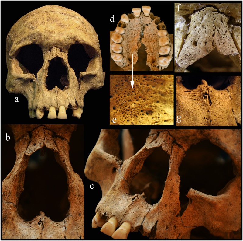
A part of the basal and the occipital regions of the skull are missing. The postcranial bones are fragmentary and partially missing. Most of the characteristic alterations are located mainly on the facial skeleton and on the lower limb bones (Figs 4a–4g and 5a–5f).
Fig 4.
a-c: Rhinomaxillary syndrome on the skull, feature 257 S20. d-e: The hard palate is porotic. f-g: The nasal bones and the anterior nasal spine have atrophied with serious inflammation.
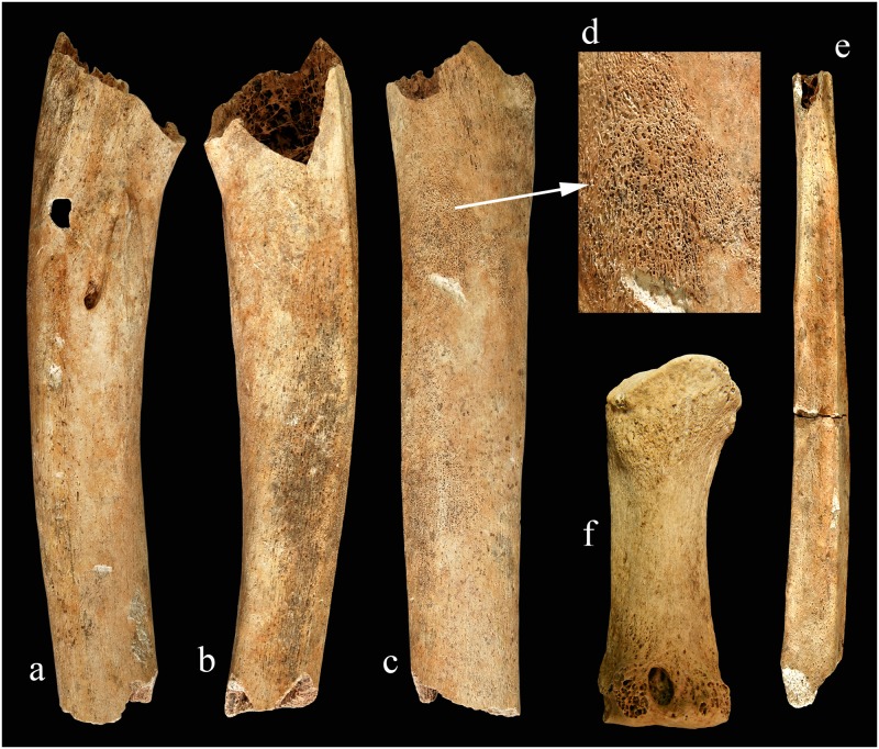
Fig 5. Changes on the extremities (a-d: right tibia e: and fibula; f: right 1st metacarpal bone), feature 257 S20.
smooth remodelling of the margin of the nasal aperture (Fig 4a–4c);
complete resorption of the anterior nasal spine (Fig 4a–4c and 4g) and the vomer;
serious inflammation, periostitis and resorption on the nasal bones (Fig 4f);
porosity on the frontal process, on the infratemporal region of the maxilla, on the left maxillary sinus and on the hard palate (Fig 4d and 4e);
porosity on the cortical layer of the occipital bone, near the lambdoidal suture;
pit formation on the cortical layer of the mandible below the incisors;
porotic vertebral bodies;
periostitis and hypervascularisation on the ribs;
periostitis on the right tibia and fibula with abnormal morphology of these bones (Fig 5a–5e);
periostitis on the diaphyseal part of both humeri (mainly on the left deltoid tuberosity), on the medial surface of both radii, on both ulnae distal end, on both femurs (from the distal of the lesser trochanter) and on the plantar surface of the right sided first os metacarpal with a deep cavity (Fig 5f);
cribra orbitalia on the left orbital roof;
two healed antemortem trauma in the frontal bone (see above the detailed description of the injuries–S1 Fig).
In a further four cases (feature 263 S25 –S10 Fig; feature 263 S36 –S11 Fig; feature 263 S29 –S12 Fig; feature 263 S39 –S13 Fig) we observed some bone lesions which possibly suggest the presence of leprosy infection but the diagnosis is very uncertain. We provide the detailed description of the pathological lesions of these individuals in the S2 Text.
Mycobacterium leprae and Mycobacterium tuberculosis aDNA analysis
The five macroscopically suspected leprosy cases were subjected to aDNA analysis of the human bones as described above, using three separate sets of extractions. Traditional PCR analysis was performed on the first two sets of extractions, with three primer pairs targeting an outer 129bp and inner 99bp sequence, or a one-stage 111bp sequence. Real-Time PCR analysis was used in the third set of extractions using RLEP primers and probe for an 80bp target and RepLep primers and probe for a 66bp target [16]. Unfortunately, no M. leprae ancient DNA was detected in the bone samples examined, although positive DNA extraction controls were successful. Neither was M. tuberculosis detected in the batch 1 extractions.
Differential diagnosis
In leprosy, destruction of the nasal bones, nasal septum and conchae, and the hard palate are the common complications on the skull [35,57]. Møller-Christensen [58,59] observed that in archaeological human remains infected by leprosy, the atrophy of the anterior nasal spine and the premaxillary alveolar process with or without loss of the upper incisors, the rounding and widening of the nasal aperture, destruction of the nasal septum and of the hard palate can be found. Neurotrophic alterations of the hands and feet are common bone lesions observed in advanced leprosy. Neurologic problems of the foot can lead to the destruction of the foot, with serious bone loss, severe disfigurement and loss of biomechanical function. Periostitis on the bones of the lower extremities (tibiae, fibulae and the bones of the feet) can be caused by chronic infection of the feet [35].
Some of the skeletal anomalies typical of leprosy also occur in other diseases, such as fungal, treponemal, and oral infections, maxillary sinusitis and leishmaniasis [35], also skeletal tuberculosis, brucellosis and neoplastic disease (metastatic carcinoma).
Fungal infections often cause lytic lesions in the bones. For example, cryptococcosis usually affects the cranial and occasionally other bones, however new bone formation is uncommon. Mucormycosis destroys the nasal cavity, the maxillary sinus and the hard palate, but causing only unilateral perforations. Maduromycosis is distributed in tropical and subtropical regions, while sporotrichosis occurs in humid areas [60]. In all fungal infections the bony changes are unilateral, there is only little destruction and no marginal repair or remodeling [61]. In our cases the lesions are not unilateral and some cases demonstrate a subperiosteal bone reaction. Maduromycosis is excluded due to its geographic range and sporotrichosis is excluded due to its link with high humidity.
In a treponemal disease, such as syphilis, one of the most characteristic lesions is the classical “sabre” shin of the tibiae. Gummatous lesions and a striated nodule on the tibial shaft and a rebuilt trabecular system of the tibia can also occur. Alterations in the skull (mainly in the cranial vault) are frequent and are produced by a combination of destruction and healing accompanied by osteosclerosis (caries sicca). Remodeling of the nasal aperture (including loss of the nasal spine) can occur [35,62,63]. The Abony individuals demonstrate no evidence of these treponemal diagnostic criteria such as: saber tibiae, gummatous lesions and a striated nodule on the tibial shaft, newly built spongiosa in the tibia, widespread periostitis in the axial and appendicular skeleton, irregular, thick long bones, cavitating lesions on the cranium or caries sicca in the cranial vault.
Oral infections and rhinomaxillary sinusitis can cause inflammatory changes to the rhinomaxillary region, and can lead to antemortem tooth loss and destruction of the alveolar part of the maxilla or the mandible [61]. Although in leprosy antemortem tooth loss is frequent, in the Abony cases this phenomenon was not observed. In the case of feature 257 S20, the alveolar process of the anterior part of the maxilla had started to atrophy without any serious inflammation.
Mucocutaneous leishmaniasis can also cause destructive alterations on the bones of the face, with particularly periosteal reactions around the anterior nasal spine and nasal antrum. These affect the mucosal tissues resulting in severe disfigurement of the face. However, there are no lesions on the long bones [35,61]. The alterations on the face are not known to cause destruction of the piriform aperture and the nasal spine that are important diagnostic criteria for leprosy. In the cases presented in this study the atrophy of the nasal spine and the piriform aperture is present.
Bone lesions in skeletal tuberculosis are located most frequently in the spine, tarsals, metatarsals, carpals and metacarpals, knee, hip and elbow joints. Bony manifestations of tuberculosis are well-known and include lytic lesions, vertebral collapse, gibbus, reactive bone formation, etc. The skull (cranial base, cranial vault, and the face) is a very rare area of involvement and occurs mainly in childhood. The cranial vault is the most frequent location of cranial tuberculosis, and the base of the skull is rarely involved. Among the facial bones, the maxilla, especially at the junction with the zygoma, is affected and in the zygomatic arch there may be abscess involvement of temporal bone and the mastoid process, together with a periosteal reaction. The nasal cavity may be affected in a secondary reaction [35]. The skeletal remains at the Abony site are of adults and they do not demonstrate the classic manifestations or localisations consistent with tuberculosis (lesions on spine, joints and other postcranial bones).
Brucellosis is an infectious disease in which the domestication of animals plays an important role. Skeletal involvement of this disease is rare. The most common skeletal lesion is in the spine or sacroiliac joint, long bones are relatively involved. In the spine, brucellosis does not result in collapse of the vertebral bodies or angular kyphosis. Brucellosis causes destructive lesions of the anterior vertebral bodies with reactive projections, and multifocal cavitating abscesses with perforation of the intervertebral disk [35]. The vertebrae and other postcranial bones of our cases show no such multifocal lytic lesions.
In neoplastic diseases (metastatic tumors) the bones are commonly involved. There are two types of bony alterations: osteolytic and osteoblastic. Osteolytic formations may be manifested in the skull and on the facial bones. Carcinoma of the nasal cavity causes destruction of the hard palate and the inner portion of the maxilla, whereas carcinoma of the ethmoid may destroy the frontal bone and the orbital wall [35,64]. However, in all cases the complete reactive bone is absent. These lesions, the absence of the reactive bone and the strictly localized region of the destruction differ from the paleopathology of leprosy cases.
Based on the above-mentioned diagnostic criteria, the young male from feature 257 S20 shows strong evidence for the bony manifestation of advanced leprosy (Figs 5 and 6).
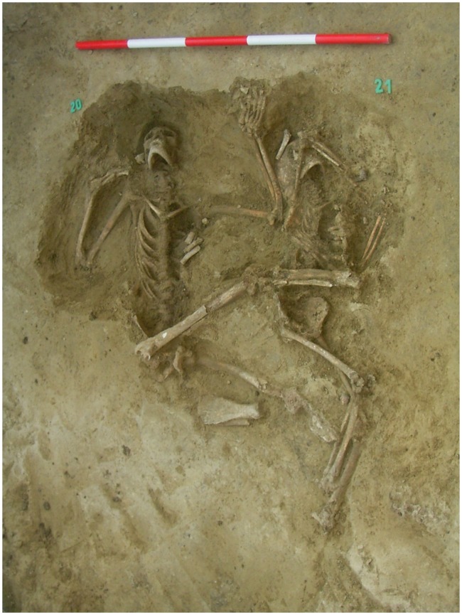
Fig 6. The young male laid on his back, who was thrown into the pit; feature 257 S20.
Based on the Modified Istanbul Protocol [65] this case shows the typical signs of leprosy. In the mass grave the male is laid on his back and he was thrown into the pit with no sign of any burial rite (Fig 6).
In a further two cases (feature 263 S36 –S11 Fig; feature 263 S39 –S13 Fig) there is a suspicion of a leprosy infection. Although other infections cannot be excluded, using the Modified Istanbul Protocol, the paleopathology is highly consistent with leprosy [65]. On the skull of feature 263 S39 the inflammation in the premaxillary area may be caused by an oral infection originating from three abscesses localized to the roots of the upper incisors (S13b Fig).
Discussion
The archaeological circumstances of the Abony site are significantly different from the pits and settlements that are known from the early, Protoboleráz phase of the Late Copper Age Carpathian Basin [6,7,13]. Based on these archaeological data the ritual origin of the features cannot be excluded. It was proved first, that some pits were thoroughly prepared, before throwing or burying the bodies. Secondly, all of the pits had the same shape. Moreover, the filling in the pits consisted of alternating layers of black humus and sterile loess layers, which suggests a repeated use of the pits. Finally in other pits, there was a posthole-like hollow at the bottom. In many cases systematically placed animal skulls, partial animal skeletons and intact vessels were found in these hollows [5]. The age distribution of the Abony population gave unexpected results when compared with the published paleodemographical data from the investigated region and period (Table 7).
Table 7. Percent proportion of fetuses and neonates in the population of Abony and other prehistoric series from the Carpathian Basin.
| Archaeological site | Age (A.), archaeological culture (c.) / group (g.) | Number of individuals | Number of fetuses and neonates | Fetuses and neonates (%) |
|---|---|---|---|---|
| Tiszapolgár-Basatanya* [66] | Copper A., Tiszapolgár c. | 59 | 0 | 0.0 |
| Tiszapolgár-Basatanya* [66] | Copper A., Bodrogkeresztúr c. | 87 | 0 | 0.0 |
| Hejőkürt-Lidl Logisztikai Központ [67] | Neolithic, Eastern Linear Pottery c. (ALPC/ELPC) | 38 | 0 | 0.0 |
| Balatonszárszó-Kis-erdei dűlő [68] | Neolithic, Linear Pottery c. (LPC) | 43 | 0 | 0.0 |
| Nitra-H. Krškany [69] | Neolithic, LPC | 73 | 1 | 1.4 |
| Aszód-Papi földek [70] | Neolithic, Lengyel c. | 197 | 5 | 2.5 |
| Mezőcsát, Bodrogkeresztúr, Tiszapolgár-Csőszhalom, Tiszapolgár, Tiszapolgár-Basatanya [71] | Copper A., Bodrogkeresztúr c., Baden c. | 71 | 2 | 2.8 |
| Budakalász-Luppa csárda [72] | Copper A., Baden c. | 406 | 13 | 3.2 |
| Polgár-Ferenci hát [73] | Neolithic, ALPC | 120 | 4 | 3.3 |
| Hajdúnánás-Eszlári út [73] | Neolithic, ALPC | 47 | 2 | 4.3 |
| Mórágy B.1 [74] | Neolithic, Lengyeli c. | 82 | 5 | 6.1 |
| Balatonőszöd-Temetői dűlő [75,76] | Copper A., Baden c. | 26 | 2 | 7.7 |
| Alsónémedi [25] | Copper A., Baden c. | 44 | 4 | 9.1 |
| Mezőkövesd-Mocsolyás [73] | Neolithic, Szatmár g. | 28 | 3 | 10.7 |
| Jánoshida-Berek [77] | Bronze A., Tumulus c. | 165 | 22 | 13.3 |
| Abony-Turjányos dűlő (present study) | Copper A., Protoboleráz horizon | 48 | 16 | 33.3 |
*anthropological results of János Nemeskéri were cited by I. B. Kutzián [66].
The ratio of the newborns and fetuses (0–1 years old children, 33,3%) is extremely high. We compared this result with the data from the anthropological literature (Table 7) involving those series (Abony, Alsónémedi, Budakalász, Jánoshida, Mórágy, Polgár-Ferenci hát) where the number of the individuals in this age group was more than three.
Statistical analysis including Chi-square and Fisher’s exact test were undertaken to determine the differences in the ratio of 0–1 years old in these populations. The ratio of the newborns and fetuses demonstrated a significant difference between the compared populations (χ2 = 51.949 DF = 5 two tailed p<0.001; Fisher’s exact test: 41.659, two tailed p value < 0.001). In a pairwise comparison with the Boneferroni correction, the proportion of the 0–1 years old age goup from the Abony population differed significantly from Polgár-Ferenci hát and Budakalász (p < 0.001). There were no significant differences (p < 0.001) with the Mórágy (p = 0.015) Alsónémedi (p = 0.285) and Jánoshida (p = 0.180) populations.
We present a high number of ante-mortem healed trauma cases in the Abony population (S1 Text). The skull injuries are typical of the use of blunt or round-edged objects, with only a small number apparently caused by wedges, triangular shaped weapons or tools. The depth of the wounds is minimal and only the external surface and the diploë of the skull are affected, suggesting that these were caused by minimal force. Most of them have healed well, with only a few cases showing signs of inflammation. These skull wounds indicate interpersonal violence among the members of the community, rather than warfare. The observed fractures of the long bones are typical of the lifestyle or may also be the result of interpersonal violence.
The observed peri-mortem trauma may have occurred as a result of intra- not inter-group violence. In these cases the ritual activity cannot also be excluded. In previous studies more cases of peri-mortem trauma have been published from the Neolithic and Copper Age [78,79], but based on our recent knowledge there is no other example in the prehistorical Europe for human bone being used as a weapon.
The detailed paleopathological observations suggest that the Copper Age population of Abony suffered from one or more serious infectious diseases, including leprosy. Among the palaeopathologies that suggest infection are endocranial lesions, especially on the occipital bone and periostitis on the long bones, which have occurred in a high proportion of bones in children.
Earlier studies have stated that the fetuses can become infected with M. leprae during pregnancy [80,81]. However, cord blood IgA is significantly increased in babies of mothers with lepromatous leprosy (LL) and IgA anti-M. leprae antibodies are present in 30% of cord sera of babies of mothers with active LL [82]. These children have a lower birth weight than the average and suffer from fetal distress [55,83,84]. Moreover, children born to mothers with leprosy are at increased risk of developing leprosy. The time it takes to develop varies according to different authors, some stating that leprosy develops by early childhood, around 2 to 5 years old or by puberty [82,85,86]. There is limited evidence of congenital or peri-natal leprosy, with the suggestion that this is often self-healing [82]. However one case of an infant aged 3–4 months with intra-cranial leprosy pathology has been described in Byzantine Turkey, suggesting there was active infection [86].
In the advanced stage of leprosy typical bone alterations (so-called facies leprosa, rhinomaxillary syndrome, lesions on the bones of the hand and feet, and especially penciling of the phalanges) can be found [58,87]. In the Abony material such typical bone alterations were recognized in one individual (Figs 4 and 5). In the case of further four individuals the observed lesions suggest the possibility of leprosy infection. Although the results of the aDNA analysis did not confirm the presence of M. leprae, this is not proof of absence. DNA is a relatively fragile molecule and its persistence depends on the local environmental conditions of the skeletal remains [45,88,89]. Initial biomolecular analyses were performed on the Abony samples to detect the possible presence of M. leprae specific mycolic acids and proteins [90,91], but the data require confirmation.
A combination of technologies can help elucidate the origin of bacterial pathogens, their distribution and spread in human populations. This information is relevant today as leprosy is still a global problem. Descriptions of bones with typical leprosy palaeopathology from the pre-historical period have been known for decades.
Written descriptions of alterations consistent with leprosy can be found in ancient Chinese scrolls that refer back to 1122 BC [92]. M. leprae is an obligate pathogen with no known environmental reservoir. Therefore, its spread around the world is linked to human migrations. According to one opinion, leprosy originated in India and the soldiers of Alexander the Great brought it into the ancient Mediterranean World after the 4th century BC [33,93]. Another suggestion is that the disease evolved in Africa [94], which is supported by M. leprae genomic data [95,96]. Monot et al. [95] presumed that all contemporary strains of leprosy can be originated to a single clone and the analysis of single nucleotide polymorphisms (SNPs) demonstrated possible origin and dispersal routes through time. They suggested that the ancestral strain of M. leprae could be either SNP1 or, more likely, SNP2. According to their opinion it is possible that M. leprae (SNP 2) appeared first in East Africa or in the Near East, with SNP 1 evolving from SNP 2, and then spreading to Asia. SNP 3 also appears to have evolved from SNP2 and was spread to Europe and the Middle East via human migrations associated with the ancient Silk Road. The Americas acquired leprosy via population movements from Europe to the Americas. Later SNP 4 arose in West Africa and thence to the Caribbean via the slave trade. Their alternative suggestion was that SNP 1 appeared in Asia, SNP 2 developed from SNP 1, SNP 3 evolved from SNP 2 and SNP 4 originated from SNP 3. In a later study, Monot et al. [96] found new evidence for 16 SNP subtypes. They suggested that there were two main routes for the spread of leprosy, a northern and a southern route. The northern route brought SNP 3K from the Mediterranean and Turkey, along the Silk Road, to Iran, China and East Asia. The southern route brought SNP 1 from India to Indonesia and the Philippines.
The possible appearance of leprosy in the Copper Age in the Carpathian Basin could be due to the human migrations that originated from the Near East. Earlier physical anthropological papers [97–99] and archaeogenetical papers [100–102] give strong support for this migration route from the Near East to the Balkan penninsula and to the Carpathian Basin in the Early Neolithic (approx. 6000 BC).
The paleopathological evidence demonstrates that leprosy was present in the Old World in prehistory. A possible leprosy case was described from the Anatolian Bronze Age (2700–2300 BC) [103] and from Balatal, Rajastan in India from the late Indus civilisation (2500–1700 BC) [61]. Another ancient putative leprosy case in Western Europe, a child with slight rhinomaxillary changes [104], was found in Dryburn Bridge, Scotland (2300–2000 BC). In Iron Age Italy at Casalecchio di Reno, Bologna, Mariotti and colleagues [105] described a skeleton (4th-3rd century BC) with typical morphological bone signs of leprosy. Leprosy cases also have been found in Uzbekistan, dating to the 1st-4th century AD [106], in 1st century Israel [107] and at the Dakhleh Oasis in Roman Egypt, dated to the early-mid 4th century AD [108]. Further cases of lepromatous leprosy have been described from mummified remains in early Christian Nubia [109] and the Byzantine period [86,110].
In Hungary the earliest written source about leprosy dates to 1082 AD[111] and Pálfi described the first leprosy case recognized by skeletal remains [112]. Subsequently an ancient M. leprae case from the Avar Period, dating to the 6-8th century AD, was described [113]. In recent years, many more leprosy cases have been diagnosed and, by using molecular analysis, have provided unequivocal evidence of this disease in Medieval Hungary. Most of these leprosy cases dated from the 7th-9th and 10th-11th centuries AD [16]. In Western Europe, the peak of morbidity was in the 12th-13th century. In the late Middle Ages there were about 19,000 leprosaria (dwellings to segregate individuals with leprosy) throughout the continent, which indicates both the increasing numbers of those infected and the fear of contagion [33,34]. Leprosy was endemic in Europe until the 16th century, but today cases from the indigenous population have almost completely disappeared [114,115].
Conclusions
Based on the appearance and frequency of healed ante- and peri-mortem trauma we can conclude that inter-personal (intra-group) violence was characteristic in the Abony Late Copper Age population. However other traces of violence were observed on the bones that appear not to have been caused by warfare or inter-group violence. Mass graves are usually associated with large epidemics. In spite of this, there were no signs of any epidemic that may have caused the bony alterations that were observed. There were layers between the human bodies which means these individuals had not died at the same time. The appearance of peri-mortem trauma is also evidence that supports the absence of an epidemic.
Based on recent results there is no way to determine the potential connections between the cases of diagnosed leprosy and the special burial circumstances.
The morphological diagnosed leprosy case from the Abony site, dated to the Late Copper Age (3780–3650 cal BC), is more than one thousand years older than the earliest known case from the Anatolian region [103]. This result significantly modifies our recent knowledge about the time and geographic spread of this specific infectious disease. These observations may stimulate other researchers to re-analyse the bioarchaeological human collections for similar findings that can provide more ancient osteological evidences for the presence of leprosy in the Old World.
Supporting information
(DOCX)
(DOCX)
(DOCX)
(DOCX)
(DOCX)
(DOCX)
The animations were made by Zsolt Réti (Institute of Archaeology, Research Centre for the Humanities, Hungarian Academy of Sciences).
(ZIP)
The animations were made by Zsolt Réti (Institute of Archaeology, Research Centre for the Humanities, Hungarian Academy of Sciences).
(RAR)
(TIF)
(TIF)
(TIF)
(TIF)
(TIF)
(JPG)
(TIF)
The fibula penetrated into the body of the 12th thoracic vertebra.
(TIF)
(TIF)
a-c: Rounded margins and horizontal vein grooves of the piriform aperture. d: Periostitis on the right tibia may be caused by trauma; feature 263 S25.
(TIF)
a-c: Possible rhinomaxillary syndrome. d: Healed periosteal lesion on the tibia. e-f: Periostitis and cavity formation on the left heel bone; feature 263 S36.
(TIF)
(TIF)
a-c: Atrophied lateral margin and inflammation of the piriform aperture and three abscesses at the roots of the upper incisors. d: ossification in the left external auditory pore; feature 263 S39.
(TIF)
Acknowledgments
We sincerely thank the reviewers of our paper for constructive criticisms, valuable comments and suggestions, which were of great help in revising the manuscript. The authors would like to thank all of those who participated in the excavation. Special thanks to Zsolt Réti (Institute of Archaeology, Research Centre for the Humanities, Hungarian Academy of Sciences) for giving his permission to use the 3D reconstruction and animation of the pits 251 and 257. TH was supported by the János Bolyai Scholarship of the Hungarian Academy of Sciences. Covering the open access publication charges for this article was provided by the Open Access Fund of the Hungarian Academy of Sciences.
Data Availability
All relevant data are within the paper and its Supporting Information files.
Funding Statement
Tamás Hajdu was supported by the János Bolyai Research Scholarship of the Hungarian Academy of Sciences. Covering the open access publication charges for this article was provided by the Open Access Fund of the Hungarian Academy of Sciences.
References
- 1.Kalicz N. Die Protoboleráz-Phase an der Grenze von zwei Epochen. In: Roman P, editor. Cernavodǎ III—Boleráz: ein vorgeschichtliches Phänomen zwischen dem Oberrhein und der Unteren Donau Symposium Mangalia/Neptun (18–24 Oktober 1999) Forschungsprogramm: Die Donau—Achse von Zivilisationen Studia Danubiana Series symposia 2. București: Institutul Român de Tracologie; 2001. pp. 385–435.
- 2.Marton T, Hansel B. Abony, Turjányos dűlő In: Kisfaludy J, editor. Régészeti Kutatások Magyarországon 2004 –Archaeological Investigations in Hungary 2004. Budapest: Kulturális Örökségvédelmi Hivatal -Magyar Nemzeti Múzeum; 2005. pp. 164–165. [Google Scholar]
- 3.Fábián S, Serlegi G. Abony, Turjányos dűlő, 1. agyagbánya In: Kisfaludi J, editor. Régészeti Kutatások Magyarországon 2006 –Archaeological Investigations in Hungary 2006. Budapest: Kulturális Örökségvédelmi Hivatal—Magyar Nemzeti Múzeum; 2007. p. 149. [Google Scholar]
- 4.Fábián S, Serlegi G. Abony, Turjányos dűlő In: Kisfaludy J, editor. Régészeti Kutatások Magyarországon 2007 –Archaeological Investigations in Hungary 2007. Budapest: Kulturális Örökségvédelmi Hivatal—Magyar Nemzeti Múzeum; 2008. p. 149. [Google Scholar]
- 5.Fábián S, Serlegi G, Daróczi-Szabó M. Burial, mass grave or sacrifice? Begräbnis, Massengrab oder Opferstelle? Salzmünde—rule or exception? Interdisciplinary Symposium 18th–20th of October 2012 in Halle (Saale). Halle: Tagungen des Landesmuseums für Vorgeschichte; 2017. p. in press.
- 6.Nevizánsky G. Grabfunde und Überbauerscheinungen der Träger der Badener Kultur im zentralen Gebiet des Karpatenbeckens. Slov Arch. 1985;33: 249–272. [Google Scholar]
- 7.Sachsse C. Unterzuchungen zu den Bestattungssitten der Badener kultur Universitätforschungen Zur prähistorischen Archäeologie 179. Bonn: Verlag Dr. Rudolf Habelt BMBH; 2010. [Google Scholar]
- 8.Serlegi G. Tikos-Homokgödrök In: Kisfaludy J, editor. Régészeti Kutatások Magyarországon 2003 –Archaeological Investigations in Hungary 2003. Budapest: Kulturális Örökségvédelmi Hivatal—Magyar Nemzeti Múzeum; 2004. pp. 301–302. [Google Scholar]
- 9.Honti S, Németh P. Balatonszemes-Szemesi-berek In: Honti S, Belényessy K, Gallina Z, Kiss V, Kulcsár G, Marton T, Nagy Á, Németh PG, Oross K, Sebők K, Somogyi K. A tervezett M7-es autópálya Somogy megyei szakaszán 2000-2001-ben végzett megelőző régészeti feltárások. Előzet. Somogyi Múzeumok Közleményei. 2002;15: 6–9. https://library.hungaricana.hu/hu/view/MEGY_SOMO_Smk_15/?pg=4&layout=s [Google Scholar]
- 10.Horváth T. The prehistoric settlement at Balatonőszöd-Temetői-dűlő Varia Archaeologica Hungarica 29. Budapest: Magyar Tudományos Akadémia Régészeti Intézete; 2014. [Google Scholar]
- 11.Honti S. Rézkori temetkezés Balatonbogláron (Ein Grab aus der Kupferzeit von Balatonboglár). Somogyi Múzeumok Közleményei. 1981;4: 25–38. [Google Scholar]
- 12.Somogyi K. Előzetes jelentés a Kaposvár– 61-es elkerülő út 29. számú lelőhelyén, Kaposújlak-Várdomb-dűlőben 2002-ben végzett megelőző feltárásról. Somogyi Múzeumok Közleményei. 2004;16: 165–178. [Google Scholar]
- 13.Bondár M. A badeni kultúra kutatási helyzete Magyarországon (vázlat). Der Forschungsstand der Badener Kultur in Ungarn (Abriss). MFMÉ –Stud Arch. 2003;8: 7–30. [Google Scholar]
- 14.Donoghue HD, Gladykowska Rzeczycka J, Marcsik A, Holton J, Spigelman M. Mycobacterium leprae in archaeological samples In: Roberts C, Lewis M, Manchester K, editors. The Past and Present of Leprosy: Archaeological, historical, palaeopathological and clinical approaches British Archaeological Reports International series. Oxford: Archaeopress; 2002. pp. 271–285. [Google Scholar]
- 15.Donoghue HD, Marcsik A, Matheson C, Vernon K, Nuorala E, Molto JE, et al. Co-infection of Mycobacterium tuberculosis and Mycobacterium leprae in human archaeological samples: a possible explanation for the historical decline of leprosy. Proceedings Biol Sci. 2005;272: 389–94. doi: 10.1098/rspb.2004.2966 [DOI] [PMC free article] [PubMed] [Google Scholar]
- 16.Donoghue HD, Michael Taylor G, Marcsik A, Molnár E, Pálfi G, Pap I, et al. A migration-driven model for the historical spread of leprosy in medieval Eastern and Central Europe. Infect Genet Evol. 2015;31: 250–6. doi: 10.1016/j.meegid.2015.02.001 [DOI] [PubMed] [Google Scholar]
- 17.Csóri Z, Donoghue HD, Marcsik A. Leprosy in the 10th–13th century AD in. Eastern Hungary. Annu Roum d’Anthropologie. 2009;46: 3–11. [Google Scholar]
- 18.Pálfi G, Molnár E. The paleopathology of specific infectious diseases from Southeastern Hungary: a brief overview. Acta Biol Szeged. 2009;53: 111–116. [Google Scholar]
- 19.Evinger S, Bernert Z, Fóthi E, Wolff K, Kovári I, Marcsik A, et al. New skeletal tuberculosis cases in past populations from Western Hungary (Transdanubia). Homo. 2011;62: 165–83. doi: 10.1016/j.jchb.2011.04.001 [DOI] [PubMed] [Google Scholar]
- 20.Hajdu T, Donoghue HD, Bernert Z, Fóthi E, Kővári I, Marcsik A. A case of spinal tuberculosis from the middle ages in Transylvania (Romania). Spine (Phila Pa 1976). 2012;37: E1598–601. [DOI] [PubMed] [Google Scholar]
- 21.Chan JZ-M, Sergeant MJ, Lee OY-C, Minnikin DE, Besra GS, Pap I, et al. Metagenomic analysis of tuberculosis in a mummy. N Engl J Med. 2013;369: 289–90. doi: 10.1056/NEJMc1302295 [DOI] [PubMed] [Google Scholar]
- 22.Masson M, Molnár E, Donoghue HD, Besra GS, Minnikin DE, Wu HHT, et al. Osteological and biomolecular evidence of a 7000-year-old case of hypertrophic pulmonary osteopathy secondary to tuberculosis from neolithic hungary. PLoS One. 2013;8: e78252 doi: 10.1371/journal.pone.0078252 [DOI] [PMC free article] [PubMed] [Google Scholar]
- 23.Köhler K, Pálfi G, Molnár E, Zalai-Gaál I, Osztás A, Bánffy E, et al. A Late Neolithic Case of Pott’s Disease from Hungary. Int J Osteoarchaeol. 2014;24: 697–703. doi: 10.1002/oa.2254 [Google Scholar]
- 24.Kay GL, Sergeant MJ, Zhou Z, Chan JZ-M, Millard A, Quick J, et al. Eighteenth-century genomes show that mixed infections were common at time of peak tuberculosis in Europe. Nat Commun. 2015;6: 6717 doi: 10.1038/ncomms7717 [DOI] [PMC free article] [PubMed] [Google Scholar]
- 25.Acsádi G, Nemeskédi J. History of Human Life, Span and Mortality. Budapest: Akadémiai Kiadó; 1970. [Google Scholar]
- 26.Stloukal M, Hanáková H. Die Lange der Langsknochen altslawischer Bevölkerungen unter besonderer Berücksichtigung von Wachstumsfragen. Homo. 1978;29: 53–69. [Google Scholar]
- 27.Fazekas IG, Kósa L. Forensic fetal osteology. Budapest: Akadémiai Kiadó; 1979. [Google Scholar]
- 28.Ferembach D, Schwidetzky I, Stloukal M. Empfehlungen für die Alters- und Geschlechtsdiagnose am Skelett. Homo. 1979;30: 1–32. [Google Scholar]
- 29.Işcan MY, Loth SR, Wright RK. Age estimation from the rib by phase analysis: white males. J Forensic Sci. 1984;29: 1094–104. Available: http://www.ncbi.nlm.nih.gov/pubmed/6502109 [PubMed] [Google Scholar]
- 30.Işcan MY, Loth SR, Wright RK. Age estimation from the rib by phase analysis: white females. J Forensic Sci. 1985;30: 853–63. Available: http://www.ncbi.nlm.nih.gov/pubmed/4031812 [PubMed] [Google Scholar]
- 31.Ubelaker D. Human Skeletal Remains Excavation, Analysis, Interpretation. Washington: Taraxacum; 1989. [Google Scholar]
- 32.Bernert Z, Évinger S, Hajdu T. New data on the biological age estimation of children using bone measurements based on historical populations from the Carpathian Basin. Ann hist-nat Mus Natn Hung. 2007;99: 199–206. Available: http://publication.nhmus.hu/pdf/annHNHM/Annals_HNHM_2007_Vol_99_199.pdf [Google Scholar]
- 33.Manchester K. The archaeology of disease. Bradford: University of Bradford Press; 1983. [Google Scholar]
- 34.Aufderheide A, Rodriguez-Martin C. The Cambridge encyclopedia of human paleopathology. Cambridge: Cambridge University Press; 1998. https://books.google.hu/books/about/The_Cambridge_Encyclopedia_of_Human_Pale.html?id=qubTdDk1H3IC&redir_esc=y [Google Scholar]
- 35.Ortner D. Identification of Pathological Conditions in Human Skeletal Remains. London-New York: Academic Press; 2003. [Google Scholar]
- 36.Kelley MA, Micozzi MS. Rib lesions in chronic pulmonary tuberculosis. Am J Phys Anthropol. 1984;65: 381–6. doi: 10.1002/ajpa.1330650407 [DOI] [PubMed] [Google Scholar]
- 37.Haas CJ, Zink A, Molńar E, Szeimies U, Reischl U, Marcsik A, et al. Molecular evidence for different stages of tuberculosis in ancient bone samples from Hungary. Am J Phys Anthropol. 2000;113: 293–304. doi: 10.1002/1096-8644(200011)113:3<293::AID-AJPA2>3.0.CO;2-6 [DOI] [PubMed] [Google Scholar]
- 38.Schultz M. Paleohistopathology of bone: a new approach to the study of ancient diseases. Am J Phys Anthropol. 2001;Suppl 33: 106–47. Available: http://www.ncbi.nlm.nih.gov/pubmed/11786993 [DOI] [PubMed] [Google Scholar]
- 39.Hershkovitz I, Greenwald CM, Latimer B, Jellema LM, Wish-Baratz S, Eshed V, et al. Serpens endocrania symmetrica (SES): a new term and a possible clue for identifying intrathoracic disease in skeletal populations. Am J Phys Anthropol. 2002;118: 201–16. doi: 10.1002/ajpa.10077 [DOI] [PubMed] [Google Scholar]
- 40.Maczel M. „On the traces of tuberculosis”. Diagnostic criteria of tuberculous affection of the human skeleton and their application in Hungarian and French anthropological series. University of la Méditerranée aix Marseille II Faculty of Medicine, Marseille.—Department of Anthropology, University of Szeged. 2003.
- 41.Stuart-Macadam P. Nutritional deficiency diseases: A survey of scurvy, rickets, and iron deficiency anemia In: Isçan M, Kennedy K, editors. Reconstruction of life from the skeleton. New York: Wiley-Liss; 1989. pp. 201–222. [Google Scholar]
- 42.Nathan H, Haas N. “Cribra orbitalia”. A bone condition of the orbit of unknown nature. Anatomical study with etiological considerations. Isr J Med Sci. 2: 171–91. Available: http://www.ncbi.nlm.nih.gov/pubmed/5912551 [PubMed] [Google Scholar]
- 43.Lovell N. Trauma Analysis in Paleopathology. Yrbk Phys Anthr. 1997;40: 139–170. [Google Scholar]
- 44.Rodríguez-Martín C. Identification and Differential Diagnosis of Traumatic Lesions of the Skeleton Forensic Anthropology and Medicine. Totowa, NJ: Humana Press; pp. 197–221. doi: 10.1007/978-1-59745-099-7_8 [Google Scholar]
- 45.O’Rourke DH, Hayes MG, Carlyle SW. Ancient DNA Studies in Physical Anthropology. Annu Rev Anthropol. 2000;29: 217–242. doi: 10.1146/annurev.anthro.29.1.217 [Google Scholar]
- 46.Poinar HN, Hofreiter M, Spaulding WG, Martin PS, Stankiewicz BA, Bland H, et al. Molecular coproscopy: dung and diet of the extinct ground sloth Nothrotheriops shastensis. Science. 1998;281: 402–6. Available: http://www.ncbi.nlm.nih.gov/pubmed/9665881 [DOI] [PubMed] [Google Scholar]
- 47.Donoghue HD, Holton J, Spigelman M. PCR primers that can detect low levels of Mycobacterium leprae DNA. J Med Microbiol. 2001;50: 177–82. doi: 10.1099/0022-1317-50-2-177 [DOI] [PubMed] [Google Scholar]
- 48.Taylor GM, Watson CL, Bouman AS, Lockwood DN M S. Variable nucleotide tandem repeat (VNTR) typing of two palaeopathological cases of lepromatous leprosy from Mediaeval England. J Archaeol Sci. 2006;33: 1569–1579. doi: 10.1016/j.jas.2006.02.008 [Google Scholar]
- 49.Abu Al-Soud W, Rådström P. Effects of amplification facilitators on diagnostic PCR in the presence of blood, feces, and meat. J Clin Microbiol. 2000;38: 4463–70. Available: http://www.ncbi.nlm.nih.gov/pubmed/11101581 [DOI] [PMC free article] [PubMed] [Google Scholar]
- 50.Forbes BA, Hicks KE. Substances interfering with direct detection of Mycobacterium tuberculosis in clinical specimens by PCR: effects of bovine serum albumin. J Clin Microbiol. 1996;34: 2125–8. Available: http://www.ncbi.nlm.nih.gov/pubmed/8862570 [DOI] [PMC free article] [PubMed] [Google Scholar]
- 51.Taylor GM, Murphy E, Hopkins R, Rutland P, Chistov Y. First report of Mycobacterium bovis DNA in human remains from the Iron Age. Microbiology. 2007;153: 1243–9. doi: 10.1099/mic.0.2006/002154-0 [DOI] [PubMed] [Google Scholar]
- 52.Köhler K, Marcsik A, Kondor K, Hajdu T. The results of the physical anthropological study of the skeletal remains from the Late Copper Age mass graves at the site of Abony, Turjányos-dűlő, Hungary. Begräbnis, Massengrab oder Opferstelle? Salzmünde—rule or exception? Interdisciplinary Symposium 18th– 20th of October 2012 in Halle (Saale). Halle: Tagungen des Landesmuseums für Vorgeschichte; 2017. pp. 209–220.
- 53.Weston DA. Investigating the specificity of periosteal reactions in pathology museum specimens. Am J Phys Anthropol. 2008;137: 48–59. doi: 10.1002/ajpa.20839 [DOI] [PubMed] [Google Scholar]
- 54.Weston DA. Brief communication: Paleohistopathological analysis of pathology museum specimens: can periosteal reaction microstructure explain lesion etiology? Am J Phys Anthropol. 2009;140: 186–93. doi: 10.1002/ajpa.21081 [DOI] [PubMed] [Google Scholar]
- 55.Lewis ME. Endocranial lesions in non-adult skeletons: understanding their aetiology. Int J Osteoarchaeol. 2004;14: 82–97. doi: 10.1002/oa.713 [Google Scholar]
- 56.Marcsik A. Egy csontelváltozás feltételezett aetiológiája. Anthropol Kozl. 1975;19: 47–53. [Google Scholar]
- 57.Job CK, Karat AB, Karat S. The histopathological appearance of leprous rhinitis and pathogenesis of septal perforation in leprosy. J Laryngol Otol. 1966;80: 718–32. Available: http://www.ncbi.nlm.nih.gov/pubmed/5940434 [DOI] [PubMed] [Google Scholar]
- 58.Møller-Christensen V. Ten lepers from Nestved in Denmark. A study of skeletons from a Medieval Danish Leper Hospital. Copenhagen: Danish Science Press; 1953. [Google Scholar]
- 59.Møller-Christensen V. Bone Changes in Leprosy. Copenhagen: Munksgaard; 1961. [Google Scholar]
- 60.Belcastro MG, Mariotti V, Facchini F, Dutour O. Leprosy in a skeleton from the 7th century necropolis of Vicenne-Campochiaro (Molise, Italy). Int J Osteoarchaeol. 2005;15: 431–448. doi: 10.1002/oa.799 [Google Scholar]
- 61.Robbins G, Tripathy VM, Misra VN, Mohanty RK, Shinde VS, Gray KM, et al. Ancient skeletal evidence for leprosy in India (2000 B.C.). PLoS One. 2009;4: e5669 doi: 10.1371/journal.pone.0005669 [DOI] [PMC free article] [PubMed] [Google Scholar]
- 62.Rogers J, Waldron T. Infections in palaeopathology: the basis of classification according to most probable cause. J Archaeol Sci. 1989;16: 611–625. doi: 10.1016/0305-4403(89)90026-5 [Google Scholar]
- 63.Smith MO, Betsinger TK, Williams LL. Differential visibility of treponemal disease in pre-Columbian stratified societies: does rank matter? Am J Phys Anthropol. 2011;144: 185–95. doi: 10.1002/ajpa.21381 [DOI] [PubMed] [Google Scholar]
- 64.Molnár E, Schultz M, Schmidt-Schultz TH, Marcsik A, Buczkó K, Zádori P, et al. Rare Case of an Ancient Craniofacial Osteosarcoma with Probable Surgical Intervention. Pathol Oncol Res. 2017;23: 583–587. doi: 10.1007/s12253-016-0153-7 [DOI] [PubMed] [Google Scholar]
- 65.Appleby J, Thomas R, Buikstra J. Increasing confidence in paleopathological diagnosis—Application of the Istanbul terminological framework. Int J Paleopathol. 2015;8: 19–21. doi: 10.1016/j.ijpp.2014.07.003 [DOI] [PubMed] [Google Scholar]
- 66.Kutzián B. The Copper Age cemetery of Tiszapolgár-Basatanya Arch Hung. 42 Budapest: Akadémiai Kiadó; 1963. [Google Scholar]
- 67.Kővári I, Szathmáry L. Neolit csontvázleletek vizsgálata Északkelet-Magyarországról (előzetes tanulmány). In: Isépy I, Korsós Z, Pap I, editors. IV Kárpát-medencei Biológiai szimpózium Előadások összefoglalói. Magyar Biológiai Társaság—Magyar Természettudományi Múzeum; 2005. pp. 129–133.
- 68.Zoffmann K. Anthropological analysis of the burials from the LBK settlement at Balatoszárszó-Kis-erdei dűlő. Acta Arch Hung. 2012;63: 301–316. [Google Scholar]
- 69.Pavúk J. Neolitisches Gräberfeld in Nitra. Slov Arch. 1972;20: 5–105. [Google Scholar]
- 70.Zoffmann K. A Lengyeli kultúra Aszód-Papiföldek lelőhelyéről származó embertani sorozat demográfiai és kraniometriai jellemzői (Individual demographical and craniometrical data of the Aszód-Papiföldek series dated to the Lengyel culture). Folia Anthr. 2015;14: 75–92. [Google Scholar]
- 71.Ubelaker DH, Pap I. Skeletal evidence for morbidity and mortality in Copper Age samples from northeastern Hungary. Int J Osteoarchaeol. 2009;19: 23–35. doi: 10.1002/oa.969 [Google Scholar]
- 72.Köhler K. The anthropological remains from the Budakalász Cemetery In: Bondár M, Raczky P, editors. The Copper Age cemetery of Budakalász. Budapest: Pytheas; 2009. pp. 304–364. [Google Scholar]
- 73.Zoffmann K. Kárpát-medence területéről származó neolitikus, réz-, bronz-, és vaskori antropológiai sorozatok halandósági táblái. (Adatközlés) (Life tables of anthropological series from the Neolithic, Copper, Bronze and Iron Ages from the Carpathian Basin). Folia Anthr. 2011;10: 17–57. [Google Scholar]
- 74.Zoffmann K. A Lengyeli kultúra Mórágy B.1. temetkezési csoportjának embertani ismertetése (Anthropological description of the Mórágy B.1 burial group of the Lengyel culture). A Wosinsky Mór Múzeum Évkönyve. 2004;26: 137–179. [Google Scholar]
- 75.Zoffmann K. A Badeni népesség Balatonőszöd lelőhelyről való embertani leleteinek ismertetése (Anthropological study on the Baden population of the Balatonőszöd site). Somogyi Múzeumok Közleményei. 2004;16: 111–125. [Google Scholar]
- 76.Köhler K. A lelőhely késő rézkori embertani leleteinek antropológiai vizsgálata In: Horváth T, editor. Balatonőszöd–Temetői dűlő őskori településrészei: a középső rézkori, késő rézkori és kora bronzkori települések. Budapest: Magyar Tudományos Akadémia Régészeti Intézete; 2012. pp. 239–270. [Google Scholar]
- 77.Hajdu T. A késő-bronzkori halomsíros kultúra Jánoshida-Berek lelőhelyen feltárt temetőjének embertani vizsgálata. (Anthropological examination of the Late Bronze Age cemetery of Tumulus culture from Jánoshida-Berek). Anthropol Kozl. 2008;49: 65–82. Available: http://www.mbt-biologia.hu/gen/pro/mod/let/let_fajl_kiiras.php?i_faj_azo=971 [Google Scholar]
- 78.Zoffmann K. Anthropological material from a neolithic common grave found at Esztergályhorváti (Lengyel culture, Hungary). Folia Anthr. 2007;6: 53–60. [Google Scholar]
- 79.Stuchliková J, Stuchlik S, Stloukal M. Ein Veterov-Massenbegrabnis ausVelké Pavlovicé. Anthropologie. 1985;23: 51–68. [Google Scholar]
- 80.Brubaker ML, Meyers WM, Bourland J. Leprosy in children one year of age and under. Int J Lepr Other Mycobact Dis. 1985;53: 517–23. Available: http://www.ncbi.nlm.nih.gov/pubmed/4086915 [PubMed] [Google Scholar]
- 81.Duncan ME. Leprosy in children one year of age and under. Int J Lepr Other Mycobact Dis. 1986;54: 646 Available: http://www.ncbi.nlm.nih.gov/pubmed/3819534 [PubMed] [Google Scholar]
- 82.Duncan ME. An historical and clinical review of the interaction of leprosy and pregnancy: a cycle to be broken. Soc Sci Med. 1993;37: 457–72. Available: http://www.ncbi.nlm.nih.gov/pubmed/8211258 [DOI] [PubMed] [Google Scholar]
- 83.Lockwood DN, Sinha HH. Pregnancy and leprosy: a comprehensive literature review. Int J Lepr Other Mycobact Dis. 1999;67: 6–12. Available: http://www.ncbi.nlm.nih.gov/pubmed/10407623 [PubMed] [Google Scholar]
- 84.Fatola CO, Venyo AK-G, Venyo LK-G, Maloney DJL. Leprosy in pregnancy—a review of the literature. HAMDAN Med J. 2015;8 doi: 10.7707/hmj.320 [Google Scholar]
- 85.Böddinghaus BK, Ludwig RJ, Kaufmann R, Enzensberger R, Gies V, Kramme S, et al. Leprosy in a pregnant woman. Infection. 2007;35: 37–9. doi: 10.1007/s15010-006-5129-1 [DOI] [PubMed] [Google Scholar]
- 86.Rubini M, Erdal YS, Spigelman M, Zaio P, Donoghue HD. Paleopathological and Molecular Study on Two Cases of Ancient Childhood Leprosy from the Roman and Byzantine Empires. Int J Osteoarchaeol. 2014;24: 570–582. doi: 10.1002/oa.2242 [Google Scholar]
- 87.Brothwell D, Sandison AT. Diseases in antiquity. Springfield: Thomas; 1967. [Google Scholar]
- 88.Pääbo S, Poinar H, Serre D, Jaenicke-Despres V, Hebler J, Rohland N, et al. Genetic analyses from ancient DNA. Annu Rev Genet. 2004;38: 645–79. doi: 10.1146/annurev.genet.37.110801.143214 [DOI] [PubMed] [Google Scholar]
- 89.Lee O-C, Bull I, Molnár E, Marcsik A, Pálfi G, Donoghue H, et al. Integrated strategies for the use of lipid biomarkers in the diagnosis of ancient mycobacterial disease. In: Mitchell P, Buckberry J, editors. Proceedings of the Twelfth Annual Conference of the British Association for Biological Anthropology and Osteoarchaeology, Department of Archaeology and Anthropology University of Cambridge 2010; BAR International Series 2380. Oxford: Archaeopress; 2012. pp. 63–69.
- 90.Köhler K, Marcsik A, Donoghue H, Márk L, Hajdu T. Előzetes eredmények az Abony 36. lelőhely késő rézkori áldozati gödreiből feltárt embertani leletek vizsgálata alapján (Preliminary results based on the anthropological examination of the skeletons of the Late Copper Aged multiple burials from Abony, site. Folia Anthr. 2009;8: 41–46. [Google Scholar]
- 91.Hajdu T, Márk L, Donoghue H, Fábián S, Marton T, Serlegi G, et al. Preliminary study of biochemical analysis of osseous leprosy dated to the Late Copper Age (3700–3600 BC) mass grave in Hungary. 18th European Meeting of the Paleopathology Association 2010 23rd August 2010 Program and Abstracts. Vienna; 2010. p. 15. [Google Scholar]
- 92.Skinsnes OK, Chang PH. Understanding of leprosy in ancient China. Int J Lepr Other Mycobact Dis. 1985;53: 289–307. Available: http://www.ncbi.nlm.nih.gov/pubmed/3894541 [PubMed] [Google Scholar]
- 93.Mark S. Alexander the Great, seafaring, and the spread of leprosy. J Hist Med Allied Sci. 2002;57: 285–311. Available: http://www.ncbi.nlm.nih.gov/pubmed/12211973 [DOI] [PubMed] [Google Scholar]
- 94.Grmek M. Les maladies á l’aube de la civilisation occidentale. Paris: Payot; 1983. [Google Scholar]
- 95.Monot M, Honoré N, Garnier T, Araoz R, Coppée J-Y, Lacroix C, et al. On the origin of leprosy. Science. 2005;308: 1040–2. doi: 10.1126/science/1109759 [DOI] [PubMed] [Google Scholar]
- 96.Monot M, Honoré N, Garnier T, Zidane N, Sherafi D, Paniz-Mondolfi A, et al. Comparative genomic and phylogeographic analysis of Mycobacterium leprae. Nat Genet. 2009;41: 1282–9. doi: 10.1038/ng.477 [DOI] [PubMed] [Google Scholar]
- 97.Zoffmann K. Anthropological sketch of the prehistoric population of the Carpathian Basin. Acta Biol Szeged. 2000;44: 75–79. [Google Scholar]
- 98.Pinhasi R, von Cramon-Taubadel N. Craniometric data supports demic diffusion model for the spread of agriculture into Europe. PLoS One. 2009;4: e6747 doi: 10.1371/journal.pone.0006747 [DOI] [PMC free article] [PubMed] [Google Scholar]
- 99.von Cramon-Taubadel N, Pinhasi R. Craniometric data support a mosaic model of demic and cultural Neolithic diffusion to outlying regions of Europe. Proceedings Biol Sci. 2011;278: 2874–80. doi: 10.1098/rspb.2010.2678 [DOI] [PMC free article] [PubMed] [Google Scholar]
- 100.Chikhi L, Nichols RA, Barbujani G, Beaumont MA. Y genetic data support the Neolithic demic diffusion model. Proc Natl Acad Sci U S A. 2002;99: 11008–13. doi: 10.1073/pnas.162158799 [DOI] [PMC free article] [PubMed] [Google Scholar]
- 101.Bramanti B, Thomas MG, Haak W, Unterlaender M, Jores P, Tambets K, et al. Genetic discontinuity between local hunter-gatherers and central Europe’s first farmers. Science. 2009;326: 137–40. doi: 10.1126/science.1176869 [DOI] [PubMed] [Google Scholar]
- 102.Mathieson I, Alpaslan Roodenberg S, Posth C, Szécsényi-Nagy A, Rohland N, Mallick S, et al. The Genomic History Of Southeastern Europe. bioRxiv. 2017; Available: http://biorxiv.org/content/early/2017/05/21/135616.abstract [DOI] [PMC free article] [PubMed] [Google Scholar]
- 103.Mellink MJ, Angel JL. Excavations at Karatas-Semayuk and Elmali, Lycia, 1969. Am J Archaeol. 1970;74: 245 doi: 10.2307/503101 [Google Scholar]
- 104.Roberts C. Human remains from the cists. In: Dunwell A. Cist burials and an Iron Age settlement at Dryburn Bridge, Innerwick, East Lothian. Scottish Archaeol Internet Rep. 2007;24: 18–25. Available: http://archaeologydataservice.ac.uk/archives/view/sair/contents.cfm?vol=24 [Google Scholar]
- 105.Mariotti V, Dutour O, Belcastro MG, Facchini F, Brasili P. Probable early presence of leprosy in Europe in a Celtic skeleton of the 4th-3rd century BC (Casalecchio di Reno, Bologna, Italy). Int J Osteoarchaeol. 2005;15: 311–325. doi: 10.1002/oa.775 [Google Scholar]
- 106.Taylor GM, Blau S, Mays S, Monot M, Lee OY-C, Minnikin DE, et al. Mycobacterium leprae genotype amplified from an archaeological case of lepromatous leprosy in Central Asia. J Archaeol Sci. 2009;36: 2408–2414. doi: 10.1016/j.jas.2009.06.026 [Google Scholar]
- 107.Matheson CD, Vernon KK, Lahti A, Fratpietro R, Spigelman M, Gibson S, et al. Molecular Exploration of the First-Century Tomb of the Shroud in Akeldama, Jerusalem. O’Rourke D, editor. PLoS One. 2009;4: e8319 doi: 10.1371/journal.pone.0008319 [DOI] [PMC free article] [PubMed] [Google Scholar]
- 108.Molto J. Leprosy in Roman period skeletons from Kellis 2, Dakhleh, Egypt In: Roberts C, Lewis M, Manchester K, editors. The past and present of leprosy Archaeological, historical, paleopathological and clinical approaches BAR International Series 1054. Oxford: Archaeopress; 2002. pp. 179–192. [Google Scholar]
- 109.Elliot Smith G, Dawson W. Egyptian Mummies. London: George Allen & Unwin Ltd; 1924. [Google Scholar]
- 110.Zias J. Leprosy in the Byzantine monasteries of the Judean Desert. Korot. 1985;9. [PubMed] [Google Scholar]
- 111.Fekete L. A magyarországi ragályos és járványos kórok rövid történelme [Internet]. Debrecen; 1874. https://library.hungaricana.hu/hu/view/KlasszikusOrvosiKonyvek_064/?pg=11&layout=s
- 112.Pálfi G. The first osteoarchaeological evidence of leprosy in Hungary. Int J Osteoarchaeol. 1991;1: 99–102. doi: 10.1002/oa.1390010205 [Google Scholar]
- 113.Marcsik A, Molnár E, Ősz B. Specifikus fertőző megbetegedések csontelváltozásai történeti népességek körében. Szeged: JATEPress; 2007. [Google Scholar]
- 114.Lechat M. The paleopathology of leprosy: an overview. Int J Lepr Other Mycobact Dis. 1999;67: 460–470. [PubMed] [Google Scholar]
- 115.Schuenemann VJ, Singh P, Mendum TA, Krause-Kyora B, Jäger G, Bos KI, et al. Genome-wide comparison of medieval and modern Mycobacterium leprae. Science. 2013;341: 179–83. doi: 10.1126/science.1238286 [DOI] [PubMed] [Google Scholar]
Associated Data
This section collects any data citations, data availability statements, or supplementary materials included in this article.
Supplementary Materials
(DOCX)
(DOCX)
(DOCX)
(DOCX)
(DOCX)
(DOCX)
The animations were made by Zsolt Réti (Institute of Archaeology, Research Centre for the Humanities, Hungarian Academy of Sciences).
(ZIP)
The animations were made by Zsolt Réti (Institute of Archaeology, Research Centre for the Humanities, Hungarian Academy of Sciences).
(RAR)
(TIF)
(TIF)
(TIF)
(TIF)
(TIF)
(JPG)
(TIF)
The fibula penetrated into the body of the 12th thoracic vertebra.
(TIF)
(TIF)
a-c: Rounded margins and horizontal vein grooves of the piriform aperture. d: Periostitis on the right tibia may be caused by trauma; feature 263 S25.
(TIF)
a-c: Possible rhinomaxillary syndrome. d: Healed periosteal lesion on the tibia. e-f: Periostitis and cavity formation on the left heel bone; feature 263 S36.
(TIF)
(TIF)
a-c: Atrophied lateral margin and inflammation of the piriform aperture and three abscesses at the roots of the upper incisors. d: ossification in the left external auditory pore; feature 263 S39.
(TIF)
Data Availability Statement
All relevant data are within the paper and its Supporting Information files.


