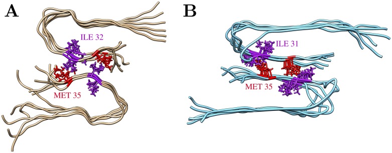
Fig 10. Comparison of the C-terminal interface between two filaments.
(A) Final structure of the AT3x∞ simulation (run2; the third loosely attached filament is omitted for clarity). (B) Twofold symmetric Aβ40 fibril structure determined by solid-state NMR spectroscopy (PDB 2LMN [30]). Interacting residues (Met35, Ile31/32) are shown in sticks.

