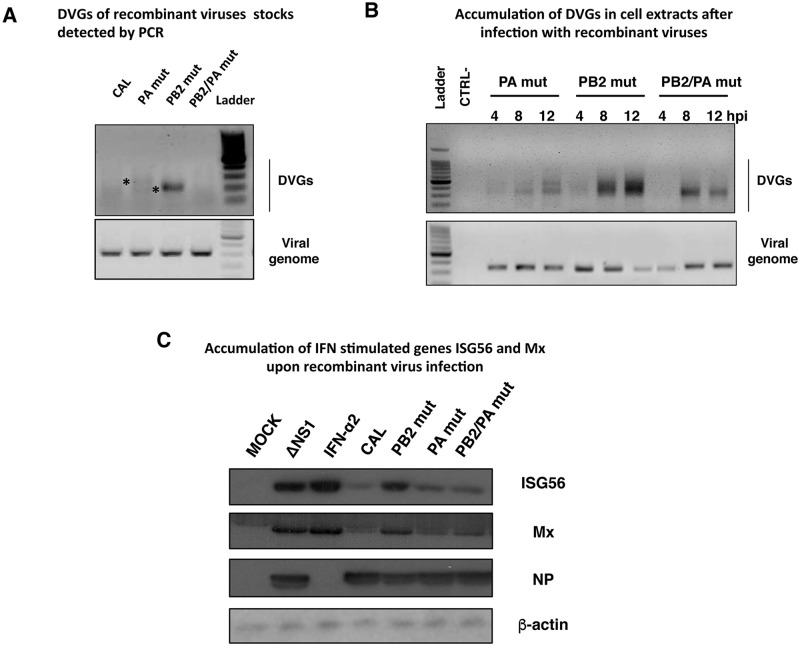
Fig 6. Different amount of defective genomes and activation of antiviral response is produced during recombinant influenza viruses infection.
(A) Detection of DVGs of PA segment in virions of CAL, PA mut, PB2 mut and PB2/PA mut recombinant viruses. Asterisks denote bands corresponding to cloned and sequenced DVGs. (B) Cultured human lung epithelial cells (A549) were infected with PB2 mut, PA mut or PB2/PA mut recombinant virus stocks at moi 1. Intracellular accumulation of DVGs was determined at indicated hours post-infection (hpi). DNA ladder size indicated in nucleotides. (C) Cultured human lung epithelial cells (A549) were infected with CAL, PB2 mut, PA mut or PB2/PA mut recombinant virus stocks at moi 1. At 16 hours post-infection (hpi), samples were used to detect the indicated proteins by Western blot. MOCK, cells treated with PBS as negative control; ΔNS1, cells infected with influenza virus lacking NS1 protein as a positive control of innate immune response activation after influenza virus infection. Virus infection was detected with antibody specific for NP, using β-actin as loading control. The experiments B and C were performed in triplicates and one representative data is shown. Quantification and significance analysis of triplicates are shown in S7 Fig.

