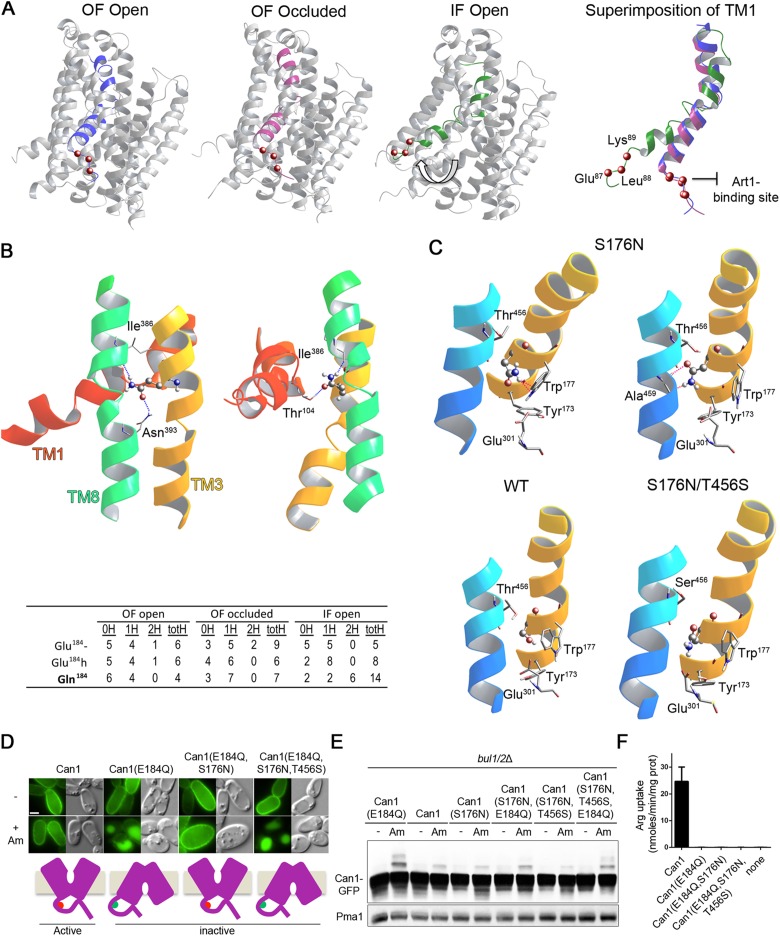
FIGURE 7:
The E184Q substitution is predicted to stabilize Can1 in an IF conformation. (A) Left, 3D models of Can1 in the OF open, OF occluded, and IF open states, highlighting a shift (white arrow) of TM1 in the IF open conformation. Right, Close-up of TM1 colored in blue, purple, and green, respectively, in the OF open, OF occluded, and IF open conformations. The location of the 87–89 sequence is shown as red balls marking the Cα position of the residues. (B) Top, view of the surroundings of Gln-184 in two 3D models of substrate-free IF open Can1(E184Q). Gln-184 is shown as balls and sticks and the residue hydrogen-bonding to it is shown as sticks. A ribbon diagram depicting neighboring residues of TM1, TM3, and TM10 is also shown. Hydrogen bonds formed by Gln-184 are shown as blue broken lines. Bottom, summary table of the analysis of H-bonds formed by residue at position 184 in structural models of Can1(E184Q) and wild-type Can1, with Glu-184 in the protonated (Glu-184h) or charged (Glu-184-) form, in the OF open, OF occluded and IF open conformations. 0H, 1H, 2H: number of Can1 models (out of 10) with no, one, or two H-bonds established by the side chain of residue 184 (in TM3) with residues of other TMs. totH: total number of H-bonds established by the side chain of residue 184 with other TMs in the 10 analyzed Can1 models. (C) Close-up view of the region encompassing residue 176 in representative OF occluded models of Can1, Can1(S176N,T456S), and Can1(S176N). In two Can1(S176N) models, pink broken lines show steric hindrance between the N176 side chain, depicted as balls and sticks, and neighboring residues, including those of the middle (W177) and distal (Y173, E301, W464) gates. Portions of TM1, TM3, and TM10 are depicted as ribbons. (D) Epifluorescence microscopy analysis of a gap1Δ can1Δ bul1/2Δ strain expressing Can1-GFP or the indicated mutant grown on Gal Pro. Glu was added for 1.5 h and then Am for 3 h. (E) immunoblots of cell extracts from the strains of D grown on Gal Pro. Glu was added for 0.5 h and then Am for 0.5 h. (F) 14C-Arg uptake measurements in a gap1Δ can1Δ strain expressing the indicated Can1 mutant. See also Supplemental Figure S5.

