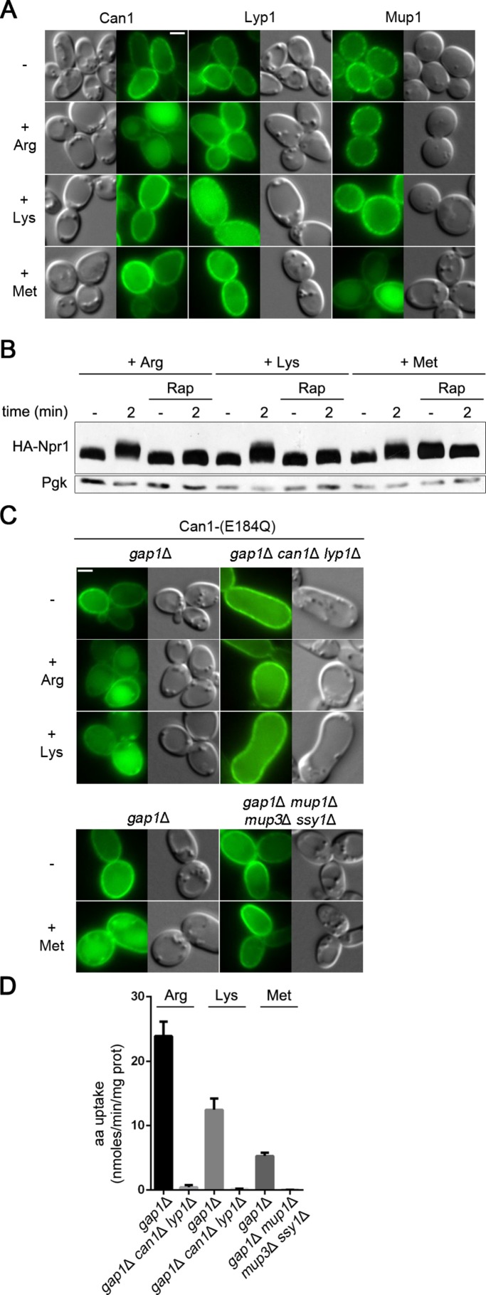
FIGURE 8:
Substrate-transport–elicited unveiling of permease cytosolic regions brings specificity to Art1-mediated ubiquitylation. (A) Epifluorescence microscopy analysis of a strain expressing Mup1-GFP and of a gap1Δ strain expressing Can1-GFP or Lyp1-GFP. For Can1-GFP, Gal Pro was used and Glu was added for 1.5 h. For Lyp1-GFP and Mup1-GFP, Glu Pro was used. Arg, Lys, or Met was added for 3 h before observation. (B) Immunoblotting of total protein extracts as in Figure 5B. Arg, Lys, or Met was added in rapamycin-treated and untreated wild-type cells. (C) Epifluorescence microscopy analysis and (D) 14C-amino acid uptake measurements on the indicated strains expressing Can1(E184Q) and grown in Gal Pro. For microscopy, Glu was added for 1.5 h and then Arg, Lys, or Met for 3 h.

