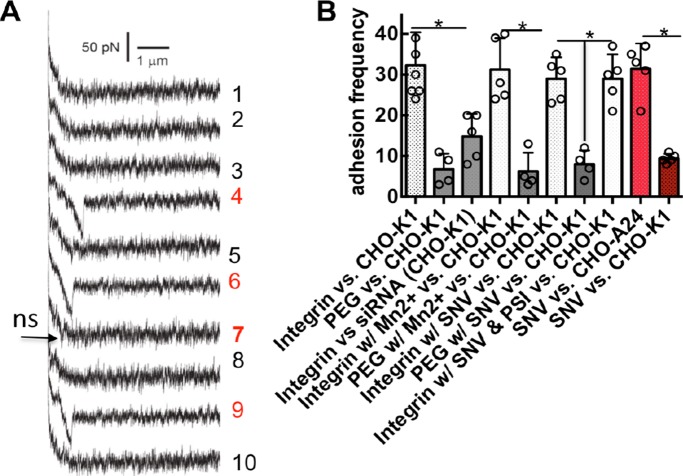
FIGURE 6:
AFM measurements of unitary αIIbβ3-RGDP2Y2R interactions performed under various conditions in WT CHO-K1 cells that are devoid of β3 integrins. Measurements of SNV-integrin (PSI) interaction measured in CHO-A24 cells stably expressing αIIbβ3 are included. (A) The measurements were performed with an adhesion frequency of ∼33%. Shown are 10 representative consecutive force-distance (retraction) traces between a cantilever tip functionalized with αIIbβ3-integrin and a CHO-K1 cell. The 4th, 6th and 9th force curves reveal unitary RGD-specific adhesive interactions. The 7th force curve shows typical nonspecific interactions, which were present with the heterobifunctional acetal-PEG27-NHS linker (PEG) only. These weak adhesions were ∼6% of the adhesion frequency events as shown in B. (B) Adhesion frequency measurements under various conditions: PEG was used to measure nonspecific interactions. siRNA refers to cells treated with P2Y2R siRNA 24 h prior to the experiment. Experiments were performed in Tyrode’s buffer (Sigma-Aldrich) containing 1 mM CaCl2, 1 mM MgCl2, 0.1% glucose, and 0.1% BSA. For Mn2+ activation, CaCl2 and MgCl2 were replaced with 2 mM MnCl2. For SNV assays, the cantilever was preincubated with fluorescently labeled neat SNVR18 or a mixture of SNVR18 and 25 µM PSI domain polypeptide to competitively block the interaction between the integrin functionalized cantilever and SNVR18. The AFM cantilever was washed before immersion into the sample chamber. Association of SNV and the integrin-functionalized AFM tip was confirmed by imaging SNV fluorescence on the AFM tip. Error bars represent SEM for greater than or equal to five separate measures such as shown in Figure 5. *p < 0.05.

