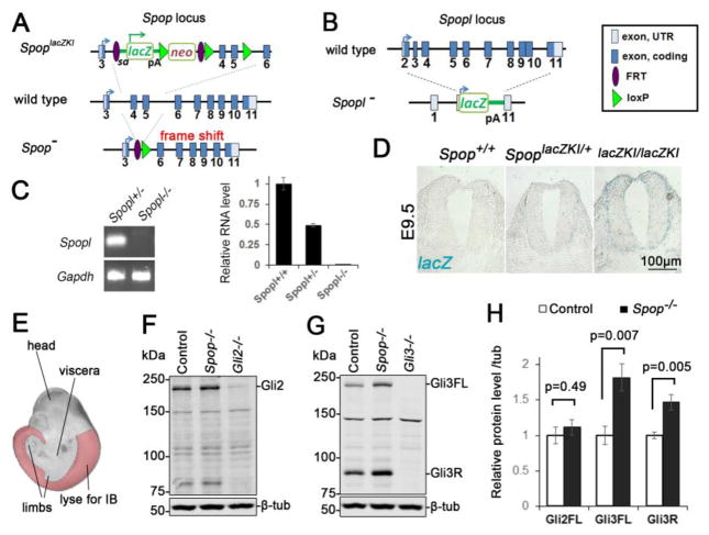
Fig. 1. A moderate increase in Gli3 protein level in the Spop mutant spinal cords.
(A) A schematic illustration of Spop loss-of-function mutant alleles. SpoplacZKI contained a lacZ and neomycin resistance cassette. SpopΔEx was generated from recombination of introduced FRT and loxP sites that deletes the 4th and 5th exons. This deletion also resulted in a frame shift that truncated the protein. (B) A schematic illustration of Spopl null mutant allele, in which a lacZ reporter replaced the entire protein-coding region of Spopl. pA: polyA signal. (C) Quantitative real time PCR showing the absence of Spopl transcript in E9.5 Spopl mutant embryos (mean ± SEM, n=3 wild type, 4 Spopl+/− and 5 Spopl−/− embryos). (D) X-gal staining of E9.5 SpoplacZKI heterozygotes and homozygotes showing Spop expression in the neural tube. (E) A schematic illustration of tissue (highlighted region) used for immunoblot in (F) and (G). The trunks of E10.5 embryos were freed of viscera and limbs to minimize the effect of Gli proteins in these tissues. (F) Immunoblots with antibodies against Gli2 and β-tubulin. (G) Immunoblots with antibodies against Gli3 and β-tubulin. (H) Quantification of (F) and (G) (mean ± SEM from n=6 embryos per group). Student’s t-test showed a significant increase in the levels of Gli3FL and Gli3R, but not Gli2 in Spop mutants.

