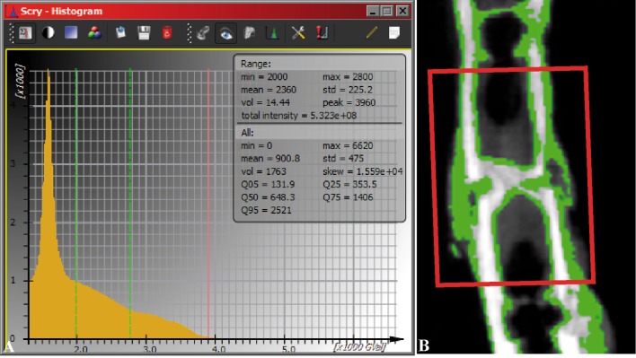
Fig. 1A–B.
The method of callus determination is shown. (A) The area between the two green lines in the histogram was assigned to callus. (B) The assigned callus is shown in green in the longitudinal bone section. The region of interest (red rectangle in illustration B) was placed at the fracture position (4 × 4 × 1.2 mm3) to calculate the callus.

