Fig. 2.

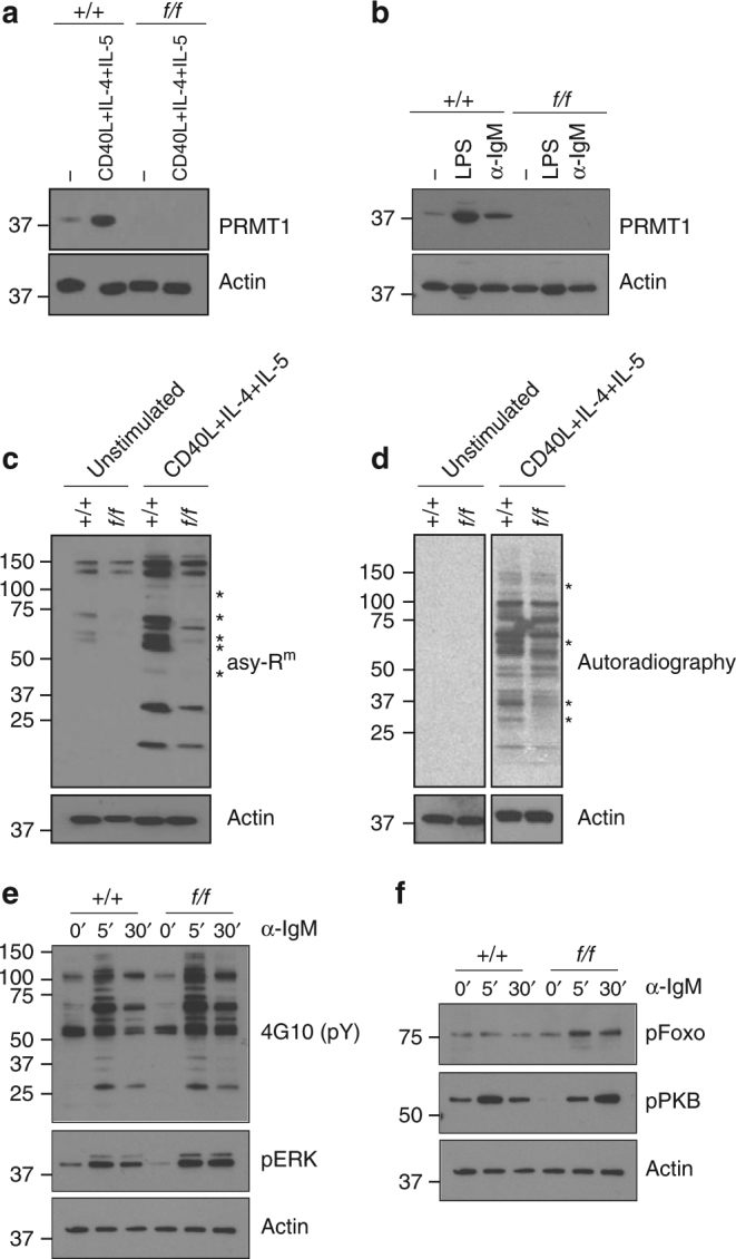
Increased PRMT1 and arginine methylated proteins in activated B cells. Immunoblot analysis of control (+/+) and Prmt1-deficient (f/f) splenic B cells for PRMT1 induction after a 2 days stimulation with CD40L and IL-4 and IL-5 or b 2 days stimulation with LPS or F(ab′)2 anti-IgM as indicated and compared to actin loading control. c Distribution of asymmetric-dimethylated arginine (asy-Rm) containing proteins in unstimulated and 2 day CD40L + IL-4 + IL-5 stimulated control (+/+) and Prmt1-deficient (f/f) splenic B cells, using specific antibody and compared to actin loading control. d Ex vivo methylation assay. Unstimulated (left panel) or day 2 CD40L + IL-4 + IL-5 activated (right panel) control (+/+) and Prmt1-deficient (f/f) B cells were grown for 3 h in the presence of L-[methyl-3H]methionine and protein synthesis inhibitors. Autoradiography of lysates reveals methylated proteins. Asterisks in c, d indicate proteins that are differentially methylated between control and Prmt1-deficient B cells. e, f B cells were activated with F(ab′)2 anti-IgM for the indicated time points. Western blot analysis of the whole-cell lysates shows the level of phospho-tyrosine (4G10), pERK (T202/Y204), e pFoxo1(pS256) and pPKB (S473) f. Actin is used as a loading control. Similar results were obtained in three independent experiments in which each sample was derived from a pool of three mice. Uncropped images of blots are presented in Supplementary Fig. 5
