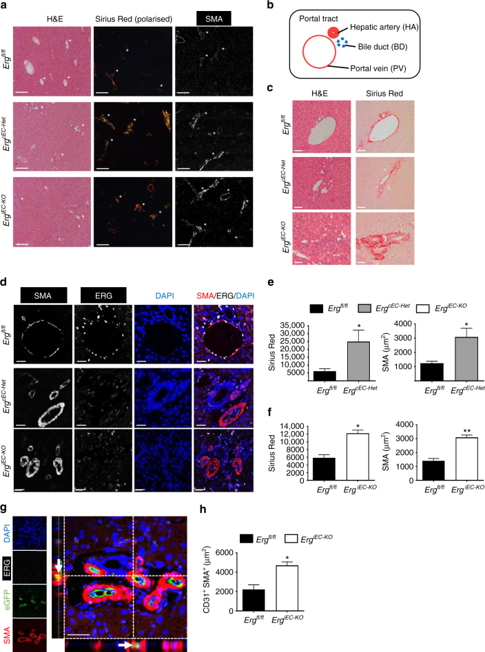
Fig. 4.
ERG-deficient mouse displays spontaneous liver fibrogenesis surrounding portal tracts. a EC-specific constitutive Erg hemi-deficient (ErgcEC-het) and inducible PDGFB-eGFP-Cre ERG flox homozygous (ErgiEC-KO) mice aged between 8 and 10 weeks were imaged by H&E, picro-sirius red and SMA (Scale bar 500 µm). Portal tract regions are identified by white asterisk with components depicted schematically in b. Portal tracts were assessed by c H&E and picro-sirius (red) (Scale bar 20 µm). d Immunofluorescence for SMA (grey scale; red in merge), ERG (white), DAPI (blue) and merged (Scale bar 20 µm). Images captured from e ErgcEC-het and f ErgiEC-KO mice were quantified for areas of Picro-sirius red positive tissue, using polarised light, and SMA expression (3 fields per mouse, n = 3). Scale bar 50 μm. g Co-localisation of SMA and eGFP expression, indicative of EndMT, was observed in ErgiEC-KO mice by immunofluorescence (double positive cell indicated by arrow in cross-section). h Quantification of CD31+SMA+ double positive cells (three fields per mouse, n = 3–4). Data were compared to Ergfl/fl littermate controls (*) by unpaired t-test. All graphical data are mean ± s.e.m., *P < 0.05, **P < 0.01

