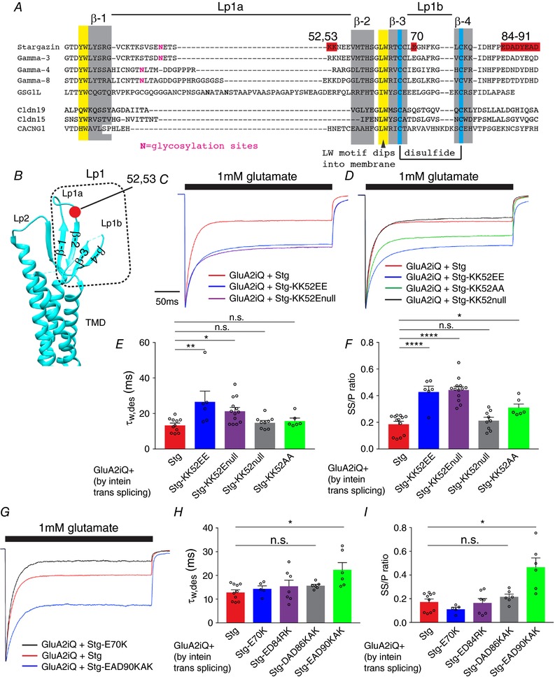
Figure 8. Gain‐of‐function mutations in Stg Lp1 and Lp2.

A, alignment of Stg, TARP γ‐3, 4, 8 and related members of the claudin family. The red residues were interrogated by mutation. The extracellular loop 1 (Lp1) is divided into Lp1a and Lp1b. Secondary structure elements and post‐translational modifications are indicated. β‐Strands (β‐1–4) and disulfide bonds are highlighted with grey and blue, respectively. B, locations that correspond to the secondary structure elements in A are mapped onto the crystal structure of claudin 19, a homologue of Stg. Lp2, extracellular loop2. The position of residues 52–53 is in red. C, D and G, representative recordings obtained from outside‐out patches in response to 1 mm glutamate application for 300 ms. The amplitude of each trace was normalized to the peak to facilitate comparison of the gating kinetics. The constructs transfected into TetON HEK cells are indicated. E, F, H and I, summary of τw,des and SS/P amplitude ratios. Individual data points are shown as open circles. Statistical significance against GluA2iQ+Stg (in red) was determined by one‐way ANOVA with post hoc Dunnett's comparison test (* P < 0.05; ** P < 0.01; **** P < 0.0001; mean ± SEM).
