Fig. 2.
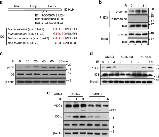
ATM- and MDC1-mediated phosphorylation of ID3 at Ser65 in response to IR. a An outline of the HLH domain and amino acid sequences including the TQ/SQ sites for human ID1, ID2, and ID3 is shown at the top of the panel. The bottom of the panel includes an alignment of ID3 HLH from four different species, demonstrating the highly conserved TQ and SQ motifs (highlighted). b HeLa cells, with or without exposure to IR, were collected at the indicated times and whole-cell lysates were subjected to immunoprecipitation using an anti-ID3 antibody followed by western blotting using anti-p-serine, anti-p-threonine, and anti-ID3 antibodies, as indicated to the right of the blot. c HeLa cells, with or without exposure to 2 Gy of IR for the indicated times, were lysed and analyzed by western blotting using anti-pSer65–ID3 and anti-ID3 antibodies. d HeLa cells were pretreated with DMSO, ATM inhibitor KU55933 (10 μM), or DNA-PK inhibitor NU7026 (5 μM) for 1 h, and then treated with or without exposure to IR for the indicated times. Cell lysates were analyzed by western blotting using anti-pSer65–ID3 and anti-ID3 antibodies. e HeLa cells transfected with either control siRNA or MDC1-specific siRNA, were treated with or without exposure to IR for the indicated times. Cell lysates were analyzed by western blotting using anti-MDC1, anti-pSer65–ID3, and anti-ID3 antibodies. Uncropped blots of this Figure accompanied by the location of molecular weight markers are shown in Supplementary Fig. 14

