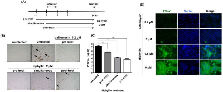
Figure 2.
Diphyllin alters cellular susceptibility against FIPV. (A) Schematic presentation of the time-of-addition assays for diphyllin treatment. 2 μM of diphyllin was added to fcwf-4 cells at three different time points relative to FIPV virus infection (MOI = 0.0001): 1 h prior to infection (pre-treat), same time as infection (simultaneous) or 1 h after infection (post-treat). After a 1 h infection period, all test cells were washed and incubated with fresh media containing 2 μM of diphyllin and incubated for 24 h. (B) Cells were then harvested for CPE observation. Virus-induced CPE are indicated by arrows. Infected cells without diphyllin treatment (untreated) or 0.2 μM bafilomycin-treated cells were used as controls. (C) Extracellular viral titers in culture supernatant were determined with plaque assays. Viral titers between each treated group and the untreated control group were compared by one-way ANOVA followed by Dunnett’s multiple comparisons test (**p < 0.01). Data in the plot present the mean ± SEM out of three replicates. (D) 0.2 μM of bafilomycin A1 or various concentrations of diphyllin (0.25, 0.5, 1, 2 μM) were added to fcwf-4 cells one hr before FIPV infection (MOI = 0.0001). Infected cells without diphyllin treatment were used as controls. After a 1-hr period of infection, cells were washed, overlaid with fresh media containing the same concentrations of diphyllin as in previous step, and incubated for another 24 hr. Fluorescence images of FITC (green), which represents expression of viral nucleocapsid protein, and nucleus (DAPI, blue) were acquired. Representative images are shown (magnification: 400×).

